

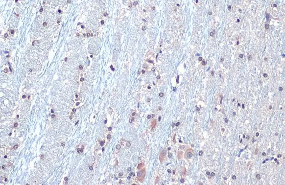
SOD1 antibody [HL1652] detects SOD1 protein at nucleus by immunohistochemical analysis. Sample: Paraffin-embedded rat brain. SOD1 stained by SOD1 antibody [HL1652] (GTX637250) diluted at 1:100. Antigen Retrieval: Citrate buffer, pH 6.0, 15 min
![SOD1 antibody [HL1652] detects SOD1 protein at cytoplasm and nucleus by immunohistochemical analysis. Sample: Paraffin-embedded human breast carcinoma. SOD1 stained by SOD1 antibody [HL1652] (GTX637250) diluted at 1:100. Antigen Retrieval: Citrate buffer, pH 6.0, 15 min](https://www.genetex.com/upload/website/prouct_img/normal/GTX637250/GTX637250_T-44753_20220826_IHC-P_22083119_636.webp "SOD1 antibody [HL1652] detects SOD1 protein at cytoplasm and nucleus by immunohistochemical analysis. Sample: Paraffin-embedded human breast carcinoma. SOD1 stained by SOD1 antibody [HL1652] (GTX637250) diluted at 1:100. Antigen Retrieval: Citrate buffer, pH 6.0, 15 min")
![Non-transfected (–) and transfected (+) 293T whole cell extracts (30 μg) were separated by 15% SDS-PAGE, and the membrane was blotted with SOD1 antibody [HL1652] (GTX637250) diluted at 1:1000. The HRP-conjugated anti-rabbit IgG antibody (GTX213110-01) was used to detect the primary antibody.](https://www.genetex.com/upload/website/prouct_img/normal/GTX637250/GTX637250_T-44753_20220916_WB_shRNA_watermark_22092119_993.webp "Non-transfected (–) and transfected (+) 293T whole cell extracts (30 μg) were separated by 15% SDS-PAGE, and the membrane was blotted with SOD1 antibody [HL1652] (GTX637250) diluted at 1:1000. The HRP-conjugated anti-rabbit IgG antibody (GTX213110-01) was used to detect the primary antibody.")
![Various tissue extracts (50 μg) were separated by 15% SDS-PAGE, and the membrane was blotted with SOD1 antibody [HL1652] (GTX637250) diluted at 1:1000. The HRP-conjugated anti-rabbit IgG antibody (GTX213110-01) was used to detect the primary antibody.](https://www.genetex.com/upload/website/prouct_img/normal/GTX637250/GTX637250_44809_20220923_WB_M_R_22100302_662.webp "Various tissue extracts (50 μg) were separated by 15% SDS-PAGE, and the membrane was blotted with SOD1 antibody [HL1652] (GTX637250) diluted at 1:1000. The HRP-conjugated anti-rabbit IgG antibody (GTX213110-01) was used to detect the primary antibody.")
![Whole cell extract (30 μg) was separated by 15% SDS-PAGE, and the membrane was blotted with SOD1 antibody [HL1652] (GTX637250) diluted at 1:1000. The HRP-conjugated anti-rabbit IgG antibody (GTX213110-01) was used to detect the primary antibody.](https://www.genetex.com/upload/website/prouct_img/normal/GTX637250/GTX637250_44809_20221223_WB_D_22122722_932.webp "Whole cell extract (30 μg) was separated by 15% SDS-PAGE, and the membrane was blotted with SOD1 antibody [HL1652] (GTX637250) diluted at 1:1000. The HRP-conjugated anti-rabbit IgG antibody (GTX213110-01) was used to detect the primary antibody.")
![SOD1 antibody [HL1652] detects SOD1 protein at cytoplasm and nucleus by immunohistochemical analysis. Sample: Paraffin-embedded dog brain. SOD1 stained by SOD1 antibody [HL1652] (GTX637250) diluted at 1:100. Antigen Retrieval: Citrate buffer, pH 6.0, 15 min](https://www.genetex.com/upload/website/prouct_img/normal/GTX637250/GTX637250_44809_20230331_IHC-P_Dog_23032819_282.webp "SOD1 antibody [HL1652] detects SOD1 protein at cytoplasm and nucleus by immunohistochemical analysis. Sample: Paraffin-embedded dog brain. SOD1 stained by SOD1 antibody [HL1652] (GTX637250) diluted at 1:100. Antigen Retrieval: Citrate buffer, pH 6.0, 15 min")
![SOD1 antibody [HL1652] detects SOD1 protein at cytoplasm and nucleus by immunohistochemical analysis. Sample: Paraffin-embedded cat brain. SOD1 stained by SOD1 antibody [HL1652] (GTX637250) diluted at 1:100. Antigen Retrieval: Citrate buffer, pH 6.0, 15 min](https://www.genetex.com/upload/website/prouct_img/normal/GTX637250/GTX637250_44809_20230331_IHC-P_Cat_23032819_338.webp "SOD1 antibody [HL1652] detects SOD1 protein at cytoplasm and nucleus by immunohistochemical analysis. Sample: Paraffin-embedded cat brain. SOD1 stained by SOD1 antibody [HL1652] (GTX637250) diluted at 1:100. Antigen Retrieval: Citrate buffer, pH 6.0, 15 min")
SOD1 antibody [HL1652] detects SOD1 protein at nucleus by immunohistochemical analysis. Sample: Paraffin-embedded rat brain. SOD1 stained by SOD1 antibody [HL1652] (GTX637250) diluted at 1:100. Antigen Retrieval: Citrate buffer, pH 6.0, 15 min
SOD1 antibody [HL1652]
GTX637250
ApplicationsWestern Blot, ImmunoHistoChemistry, ImmunoHistoChemistry Paraffin
Product group Antibodies
ReactivityCanine, Feline, Human, Mouse, Rat
TargetSOD1
Overview
- SupplierGeneTex
- Product NameSOD1 antibody [HL1652]
- Delivery Days Customer9
- Application Supplier NoteIHC-P: 1:100-1:1000. *Optimal dilutions/concentrations should be determined by the researcher.Not tested in other applications.
- ApplicationsWestern Blot, ImmunoHistoChemistry, ImmunoHistoChemistry Paraffin
- CertificationResearch Use Only
- ClonalityMonoclonal
- Clone IDHL1652
- Concentration1 mg/ml
- ConjugateUnconjugated
- Gene ID6647
- Target nameSOD1
- Target descriptionsuperoxide dismutase 1
- Target synonymsALS, ALS1, HEL-S-44, IPOA, SOD, STAHP, hSod1, homodimer, superoxide dismutase [Cu-Zn], Cu/Zn superoxide dismutase, SOD, soluble, epididymis secretory protein Li 44, indophenoloxidase A, superoxide dismutase 1, soluble, superoxide dismutase, cystolic
- HostRabbit
- IsotypeIgG
- Protein IDP00441
- Protein NameSuperoxide dismutase [Cu-Zn]
- Scientific DescriptionThe protein encoded by this gene binds copper and zinc ions and is one of two isozymes responsible for destroying free superoxide radicals in the body. The encoded isozyme is a soluble cytoplasmic protein, acting as a homodimer to convert naturally-occuring but harmful superoxide radicals to molecular oxygen and hydrogen peroxide. The other isozyme is a mitochondrial protein. Mutations in this gene have been implicated as causes of familial amyotrophic lateral sclerosis. Rare transcript variants have been reported for this gene. [provided by RefSeq, Jul 2008]
- ReactivityCanine, Feline, Human, Mouse, Rat
- Storage Instruction-20°C or -80°C,2°C to 8°C
- UNSPSC41116161
Datasheet
Related products
Product group Antibodies
SOD1 AntibodyCSB-PA004131
ApplicationsWestern Blot, ELISA
ReactivityHuman, Mouse, Rat
TargetSOD1
- SizePrice
Product group Antibodies
Anti-SOD1 AntibodyA28750
ApplicationsWestern Blot, ImmunoHistoChemistry
ReactivityHuman, Mouse, Rat
- SizePrice
Product group Antibodies

ApplicationsWestern Blot, ELISA
ReactivityHuman
TargetSOD1
- SizePrice
Product group Antibodies

Goat anti-SOD1, BiotinylatedEB07208-B
ApplicationsWestern Blot, ELISA
ReactivityHuman, Mouse, Rat
TargetSOD1
- SizePrice

Product group Antibodies
Anti-SOD1 AntibodyHPA001401
ApplicationsWestern Blot, ImmunoCytoChemistry, ImmunoHistoChemistry
ReactivityHuman, Mouse, Rat
TargetSOD1
- SizePrice
Product group Antibodies
Sod1 Polyclonal AntibodyCAC07037
ApplicationsImmunoFluorescence, Western Blot, ELISA, ImmunoHistoChemistry
ReactivityMouse
TargetSOD1
- SizePrice

Product group Antibodies
Anti-Superoxide Dismutase 1/SOD1 Antibody Picoband(r)PB9402-CARRIER-FREE
ApplicationsFlow Cytometry, ImmunoFluorescence, Western Blot, ImmunoCytoChemistry, ImmunoHistoChemistry
ReactivityHuman, Mouse, Rat
TargetSOD1
- SizePrice
![Various tissue extracts (50 μg) were separated by 15% SDS-PAGE, and the membrane was blotted with SOD1 antibody [HL1653] (GTX637251) diluted at 1:1000. The HRP-conjugated anti-rabbit IgG antibody (GTX213110-01) was used to detect the primary antibody.](https://www.genetex.com/upload/website/prouct_img/normal/GTX637251/GTX637251_T-44753_20220729_WB_M_R_22080119_341.webp)
Product group Antibodies
SOD1 antibody [HL1653]GTX637251
ApplicationsWestern Blot, ImmunoHistoChemistry, ImmunoHistoChemistry Paraffin
ReactivityCanine, Feline, Human, Mouse, Rat
TargetSOD1
- SizePrice
![SOD1 antibody [HL2033] detects SOD1 protein at cytoplasm and nucleus by immunohistochemical analysis. Sample: Paraffin-embedded mouse prostate. SOD1 stained by SOD1 antibody [HL2033] (GTX637934) diluted at 1:100. Antigen Retrieval: Citrate buffer, pH 6.0, 15 min](https://www.genetex.com/upload/website/prouct_img/normal/GTX637934/GTX637934_T-44879_20230106_IHC-P_M_23010400_601.webp)
Product group Antibodies
SOD1 antibody [HL2033]GTX637934
ApplicationsWestern Blot, ImmunoHistoChemistry, ImmunoHistoChemistry Paraffin
ReactivityCanine, Feline, Human, Mouse, Rat, Zebra Fish
TargetSOD1
- SizePrice