

Sample (30 μg of whole cell lysate) A: GL261 10% SDS PAGE GTX101506 diluted at 1:1000 The HRP-conjugated anti-rabbit IgG antibody (GTX213110-01) was used to detect the primary antibody.
. Red: Sox2 antibody (GTX101506) diluted at 1:500. Blue: DAPI")
antibody at 1:50 dilution(green) or rabbit IgG (black).")
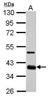
 A: A431 (GTX27909) 10% SDS PAGE GTX101506 diluted at 1:1000 The HRP-conjugated anti-rabbit IgG antibody (GTX213110-01) was used to detect the primary antibody.")
 and transfected (+) NT2D1 whole cell extracts (30 μg) were separated by 10% SDS-PAGE, and the membrane was blotted with SOX2 antibody (GTX101506) diluted at 1:1000. The HRP-conjugated anti-rabbit IgG antibody (GTX213110-01) was used to detect the primary antibody.")
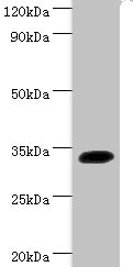
 A: Human ESC 10% SDS PAGE GTX101506 diluted at 1:1000 The HRP-conjugated anti-rabbit IgG antibody (GTX213110-01) was used to detect the primary antibody.")
 antibody at 1:100 dilution.")
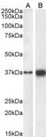
 was separated by 12% SDS-PAGE, and the membranes were blotted with SOX2 antibody (GTX101506) diluted at 1:1000 and competitor's antibody (sc-17320) diluted at 1:500. The HRP-conjugated anti-rabbit IgG antibody (GTX213110-01) was used to detect the primary antibody, and the signal was developed with Trident ECL plus-Enhanced. *The competitor is not affiliated with GeneTex and does not endorse this product.")
![Sox2 antibodies detects Sox2 proteins on embryonic mouse brain by immunohistochemical analysis. Sample:Frozen section of embryonic mouse brain (mE18.5). Green: beta III Tubulin antibody [GT886] (GTX631830) diluted at 1:2000. Red: Sox2 antibody (GTX101506) diluted at 1:250.](https://www.genetex.com/upload/website/prouct_img/normal/GTX101506/GTX101506_39911_20150625_IHC_M_w_23060100_310.webp "Sox2 antibodies detects Sox2 proteins on embryonic mouse brain by immunohistochemical analysis. Sample:Frozen section of embryonic mouse brain (mE18.5). Green: beta III Tubulin antibody [GT886] (GTX631830) diluted at 1:2000. Red: Sox2 antibody (GTX101506) diluted at 1:250.")
 diluted at 1:200. Red: beta Tubulin 3/ TUJ1 protein stained by beta Tubulin 3/ TUJ1 antibody (GTX631836) diluted at 1:200.")
Sample (30 μg of whole cell lysate) A: GL261 10% SDS PAGE GTX101506 diluted at 1:1000 The HRP-conjugated anti-rabbit IgG antibody (GTX213110-01) was used to detect the primary antibody.
SOX2 antibody
GTX101506
ApplicationsFlow Cytometry, ImmunoFluorescence, Western Blot, ImmunoCytoChemistry, ImmunoHistoChemistry, ImmunoHistoChemistry Frozen
Product group Antibodies
ReactivityHuman, Mouse
TargetSOX2
Overview
- SupplierGeneTex
- Product NameSOX2 antibody
- Delivery Days Customer9
- Application Supplier NoteWB: 1:500-1:3000. ICC/IF: 1:100-1:1000. IHC-Fr: 1:100-1:1000. FCM: 1:50-1:200. *Optimal dilutions/concentrations should be determined by the researcher.Not tested in other applications.
- ApplicationsFlow Cytometry, ImmunoFluorescence, Western Blot, ImmunoCytoChemistry, ImmunoHistoChemistry, ImmunoHistoChemistry Frozen
- CertificationResearch Use Only
- ClonalityPolyclonal
- Concentration1 mg/ml
- ConjugateUnconjugated
- Gene ID6657
- Target nameSOX2
- Target descriptionSRY-box transcription factor 2
- Target synonymsANOP3, MCOPS3, transcription factor SOX-2, SRY (sex determining region Y)-box 2, SRY-box 2, SRY-related HMG-box gene 2, sex determining region Y-box 2, transcription factor SOX2
- HostRabbit
- IsotypeIgG
- Protein IDP48431
- Protein NameTranscription factor SOX-2
- Scientific DescriptionThis intronless gene encodes a member of the SRY-related HMG-box (SOX) family of transcription factors involved in the regulation of embryonic development and in the determination of cell fate. The product of this gene is required for stem-cell maintenance in the central nervous system, and also regulates gene expression in the stomach. Mutations in this gene have been associated with optic nerve hypoplasia and with syndromic microphthalmia, a severe form of structural eye malformation. This gene lies within an intron of another gene called SOX2 overlapping transcript (SOX2OT). [provided by RefSeq]
- ReactivityHuman, Mouse
- Storage Instruction-20°C or -80°C,2°C to 8°C
- UNSPSC41116161
Datasheet
Related products
Product group Antibodies
Anti-SOX2 AntibodyA285964
ApplicationsFlow Cytometry, Western Blot, ELISA, ImmunoHistoChemistry
ReactivityHuman
- SizePrice
Product group Antibodies
Anti-SOX2 Antibody130-10236
ApplicationsWestern Blot, ELISA
TargetSOX2
- SizePrice

Product group Antibodies
Anti-SOX2 AntibodyAMAB91307
ApplicationsWestern Blot, ImmunoCytoChemistry, ImmunoHistoChemistry
ReactivityHuman, Mouse
TargetSOX2
- SizePrice

Product group Antibodies
Anti-SOX2 Antibody Picoband(r)A00105-1-CARRIER-FREE
ApplicationsWestern Blot, ELISA
ReactivityHuman, Rat
TargetSOX2
- SizePrice

Product group Antibodies
SOX2 Polyclonal AntibodyBS-23177R
ApplicationsImmunoFluorescence, Western Blot, ELISA, ImmunoCytoChemistry, ImmunoHistoChemistry, ImmunoHistoChemistry Frozen, ImmunoHistoChemistry Paraffin
ReactivityBovine, Canine, Chicken, Equine, Human, Mouse, Rat, Sheep
TargetSOX2
- SizePrice
Product group Antibodies

Goat anti-SOX2EB07378
ApplicationsFlow Cytometry, Western Blot, ELISA, ImmunoHistoChemistry
ReactivityBovine, Canine, Human, Mouse, Rat
TargetSOX2
- SizePrice
Product group Antibodies
Sox2 Polyclonal AntibodyCAC07009
ApplicationsImmunoFluorescence, Western Blot, ELISA, ImmunoHistoChemistry
ReactivityMouse
TargetSOX2
- SizePrice
Product group Antibodies
SOX2 AntibodyCSB-PA16539A0RB
ApplicationsImmunoFluorescence, Western Blot, ELISA, ImmunoHistoChemistry
ReactivityHuman, Mouse
TargetSOX2
- SizePrice
Product group Antibodies
SOX2 antibodyGTX18149
ApplicationsFlow Cytometry, ImmunoFluorescence, Western Blot, ImmunoCytoChemistry
ReactivityHuman
TargetSOX2
- SizePrice