

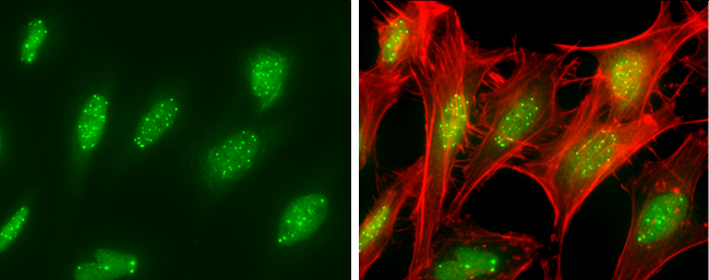
SP100 antibody detects SP100 protein at nucleus by immunofluorescent analysis. Sample: HeLa cells were fixed in 4% paraformaldehyde at RT for 15 min. Green: SP100 protein stained by SP100 antibody (GTX131569) diluted at 1:500. Red: phalloidin, a cytoskeleton marker, diluted at 1:100.
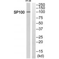
 were separated by 7.5% SDS-PAGE, and the membrane was blotted with SP100 antibody (GTX131569) diluted at 1:1000. The HRP-conjugated anti-rabbit IgG antibody (GTX213110-01) was used to detect the primary antibody.")
SP100 antibody detects SP100 protein at nucleus by immunofluorescent analysis. Sample: HeLa cells were fixed in 4% paraformaldehyde at RT for 15 min. Green: SP100 protein stained by SP100 antibody (GTX131569) diluted at 1:500. Red: phalloidin, a cytoskeleton marker, diluted at 1:100.
SP100 antibody
GTX131569
ApplicationsImmunoFluorescence, Western Blot, ImmunoCytoChemistry
Product group Antibodies
ReactivityHuman
TargetSP100
Overview
- SupplierGeneTex
- Product NameSP100 antibody
- Delivery Days Customer9
- Application Supplier NoteWB: 1:500-1:3000. ICC/IF: 1:100-1:1000. *Optimal dilutions/concentrations should be determined by the researcher.Not tested in other applications.
- ApplicationsImmunoFluorescence, Western Blot, ImmunoCytoChemistry
- CertificationResearch Use Only
- ClonalityPolyclonal
- Concentration0.68 mg/ml
- ConjugateUnconjugated
- Gene ID6672
- Target nameSP100
- Target descriptionSP100 nuclear antigen
- Target synonymslysp100b, nuclear autoantigen Sp-100, SP100-HMG nuclear autoantigen, nuclear dot-associated Sp100 protein, speckled 100 kDa
- HostRabbit
- IsotypeIgG
- Protein IDP23497
- Protein NameNuclear autoantigen Sp-100
- ReactivityHuman
- Storage Instruction-20°C or -80°C,2°C to 8°C
- UNSPSC41116161
Datasheet
Related products
Product group Antibodies
Anti-SP100 Antibody144-05851
ApplicationsWestern Blot
ReactivityHuman, Mouse
TargetSP100
- SizePrice
Product group Antibodies
Anti-SP-100 AntibodyA02051
ApplicationsImmunoFluorescence, Western Blot, ELISA, ImmunoHistoChemistry
ReactivityHuman, Mouse
TargetSP100
- SizePrice
Product group Antibodies
ApplicationsWestern Blot, ImmunoHistoChemistry
TargetSP100
- SizePrice
Product group Antibodies
SP100 AntibodyCSB-PA050127
ApplicationsWestern Blot, ELISA, ImmunoHistoChemistry
ReactivityHuman
TargetSP100
- SizePrice
Product group Antibodies

SP100 AntibodyLS-C334338
ApplicationsWestern Blot, ImmunoHistoChemistry
ReactivityHuman, Mouse
TargetSP100
- SizePrice
Product group Antibodies
SP100 antibodyGTX131570
ApplicationsImmunoFluorescence, Western Blot, ImmunoCytoChemistry
ReactivityHuman
TargetSP100
- SizePrice
Product group Antibodies
SP100 antibodyGTX32892
ApplicationsWestern Blot
ReactivityHuman, Mouse
TargetSP100
- SizePrice

Product group Antibodies
Anti-SP100 AntibodyHPA016707
ApplicationsWestern Blot, ImmunoHistoChemistry
ReactivityHuman
TargetSP100
- SizePrice