

Immunohistochemistry of paraffin-embedded Human colon cancer tissue using SPDEF Polyclonal Antibody at dilution 1:60
Immunohistochemistry of paraffin-embedded Human colon cancer tissue using SPDEF Polyclonal Antibody at dilution 1:60
SPDEF Polyclonal Antibody
E-AB-10535
ApplicationsImmunoHistoChemistry
Product group Antibodies
TargetSPDEF
Overview
- SupplierElabscience
- Product NameSPDEF Polyclonal Antibody
- Delivery Days Customer12
- ApplicationsImmunoHistoChemistry
- Applications SupplierELISA IHC
- CertificationResearch Use Only
- ClonalityPolyclonal
- Concentration1 mg/ml
- ConjugateUnconjugated
- Gene ID25803
- Target nameSPDEF
- Target descriptionSAM pointed domain containing ETS transcription factor
- Target synonymsPDEF, bA375E1.3, SAM pointed domain-containing Ets transcription factor, prostate epithelium-specific Ets transcription factor, prostate-derived Ets factor, prostate-specific Ets
- HostRabbit
- IsotypeIgG
- Protein IDO95238
- Protein NameSAM pointed domain-containing Ets transcription factor
- Scientific DescriptionThe protein encoded by this gene belongs to the ETS family of transcription factors. It is highly expressed in the prostate epithelial cells, and functions as an androgen-independent transactivator of prostate-specific antigen (PSA) promoter. Higher expression of this protein has also been reported in brain, breast, lung and ovarian tumors, compared to the corresponding normal tissues, and it shows better tumor-association than other cancer-associated molecules, making it a more suitable target for developing specific cancer therapies. Alternatively spliced transcript variants encoding different isoforms have been found for this gene.
- Storage Instruction-20°C
- UNSPSC41116161
MSDS
Related products
Product group Antibodies
SPDEF AntibodyCSB-PA022518LA01HU
ApplicationsWestern Blot, ELISA, ImmunoHistoChemistry
ReactivityHuman, Mouse
TargetSPDEF
- SizePrice
Product group Antibodies
Anti-SPDEF AntibodyA31572
ApplicationsWestern Blot, ImmunoHistoChemistry
ReactivityHuman
- SizePrice

Product group Antibodies
Anti-SPDEF AntibodyHPA055707
ApplicationsImmunoCytoChemistry
ReactivityHuman
TargetSPDEF
- SizePrice

Product group Antibodies
Anti-PSE/SPDEF Antibody Picoband(r)A04625-1-CARRIER-FREE
ApplicationsFlow Cytometry, ImmunoFluorescence, Western Blot, ELISA, ImmunoCytoChemistry
ReactivityHuman
TargetSPDEF
- SizePrice
Product group Antibodies

PDEF / SPDEF AntibodyLS-C400961
ApplicationsELISA, ImmunoHistoChemistry
ReactivityHuman, Mouse
TargetSPDEF
- SizePrice
Product group Antibodies
Spdef Polyclonal AntibodyCAC07726
ApplicationsWestern Blot, ELISA, ImmunoHistoChemistry
ReactivityMouse
TargetSPDEF
- SizePrice
Product group Antibodies
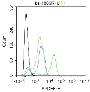
SPDEF Polyclonal AntibodyBS-1866R
ApplicationsFlow Cytometry, ImmunoFluorescence, ELISA, ImmunoCytoChemistry, ImmunoHistoChemistry, ImmunoHistoChemistry Frozen, ImmunoHistoChemistry Paraffin
ReactivityBovine, Canine, Chicken, Equine, Human, Mouse, Rabbit, Rat
TargetSPDEF
- SizePrice