

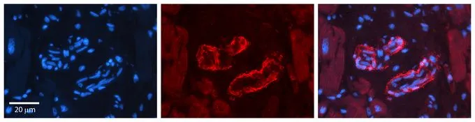
Sprouty 2 antibody Formalin Fixed Paraffin Embedded Tissue: Human heart Tissue, Observed Staining: Cytoplasmic in vasculature, Primary Antibody Concentration: 1:100, Other Working Concentrations: 1:600, Secondary Antibody: Donkey anti-Rabbit-Cy3, Secondary Antibody Concentration: 1:200 and Magnification: 20X

Sprouty 2 antibody Formalin Fixed Paraffin Embedded Tissue: Human heart Tissue, Observed Staining: Cytoplasmic in vasculature, Primary Antibody Concentration: 1:100, Other Working Concentrations: 1:600, Secondary Antibody: Donkey anti-Rabbit-Cy3, Secondary Antibody Concentration: 1:200 and Magnification: 20X
Sprouty 2 antibody
GTX33687
ApplicationsWestern Blot, ImmunoHistoChemistry, ImmunoHistoChemistry Paraffin
Product group Antibodies
ReactivityHuman
TargetSPRY2
Overview
- SupplierGeneTex
- Product NameSprouty 2 antibody
- Delivery Days Customer9
- ApplicationsWestern Blot, ImmunoHistoChemistry, ImmunoHistoChemistry Paraffin
- CertificationResearch Use Only
- ClonalityPolyclonal
- Concentration0.5-1 mg/ml
- ConjugateUnconjugated
- Gene ID10253
- Target nameSPRY2
- Target descriptionsprouty RTK signaling antagonist 2
- Target synonymsIGAN3, hSPRY2, protein sprouty homolog 2
- HostRabbit
- IsotypeIgG
- Protein IDO43597
- Protein NameProtein sprouty homolog 2
- Scientific DescriptionThis gene encodes a protein belonging to the sprouty family. The encoded protein contains a carboxyl-terminal cysteine-rich domain essential for the inhibitory activity on receptor tyrosine kinase signaling proteins and is required for growth factor stimulated translocation of the protein to membrane ruffles. In primary dermal endothelial cells this gene is transiently upregulated in response to fibroblast growth factor two. This protein is indirectly involved in the non-cell autonomous inhibitory effect on fibroblast growth factor two signaling. The protein interacts with Cas-Br-M (murine) ectropic retroviral transforming sequence, and can function as a bimodal regulator of epidermal growth factor receptor/mitogen-activated protein kinase signaling. This protein may play a role in alveoli branching during lung development as shown by a similar mouse protein. [provided by RefSeq, Jul 2008]
- ReactivityHuman
- Storage Instruction-20°C or -80°C,2°C to 8°C
- UNSPSC41116161
Datasheet
Related products
Product group Antibodies
SPRY2 AntibodyCSB-PA022622LA01HU
ApplicationsImmunoFluorescence, Western Blot, ELISA, ImmunoHistoChemistry
ReactivityHuman, Mouse
- SizePrice

Product group Antibodies
Anti-Spry-2/SPRY2 Antibody Picoband(r)A02089-2-CARRIER-FREE
ApplicationsFlow Cytometry, Western Blot, ELISA
ReactivityHuman, Mouse, Rat
TargetSPRY2
- SizePrice
Product group Antibodies

SPRY2 / Sprouty 2 AntibodyLS-C746797
ApplicationsWestern Blot
ReactivityHuman, Mouse
TargetSPRY2
- SizePrice
Product group Antibodies
Anti-SPRY2 AntibodyHPA042198
ApplicationsImmunoCytoChemistry
ReactivityHuman
TargetSPRY2
- SizePrice
Product group Antibodies
Sprouty 2 Recombinant Antibody, Biotin ConjugatedBSM-61798R-BIOTIN
ApplicationsImmunoPrecipitation, Western Blot, ImmunoHistoChemistry, ImmunoHistoChemistry Frozen, ImmunoHistoChemistry Paraffin
ReactivityHuman, Rat
TargetSPRY2
- SizePrice
Product group Antibodies
Spry2 Polyclonal AntibodyCAC07894
ApplicationsImmunoFluorescence, Western Blot, ELISA, ImmunoHistoChemistry
ReactivityMouse
- SizePrice
Product group Antibodies
Anti-SPRY2 Antibody144-65385
ApplicationsWestern Blot, ImmunoHistoChemistry
ReactivityHuman, Mouse
TargetSPRY2
- SizePrice