

srGAP3 Polyclonal Antibody
BS-3660R
ApplicationsImmunoFluorescence, Western Blot, ELISA, ImmunoCytoChemistry, ImmunoHistoChemistry, ImmunoHistoChemistry Frozen, ImmunoHistoChemistry Paraffin
Product group Antibodies
ReactivityBovine, Canine, Equine, Human, Mouse, Rabbit, Rat
TargetSRGAP3
Overview
- SupplierBioss
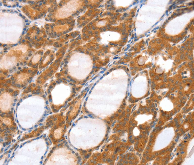
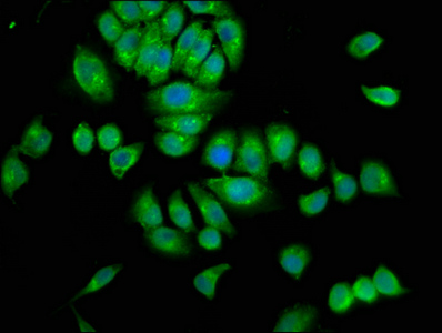
- Product NamesrGAP3 Polyclonal Antibody
- Delivery Days Customer16
- ApplicationsImmunoFluorescence, Western Blot, ELISA, ImmunoCytoChemistry, ImmunoHistoChemistry, ImmunoHistoChemistry Frozen, ImmunoHistoChemistry Paraffin
- Applications SupplierWB(1:300-5000), ELISA(1:500-1000), IHC-P(1:200-400), IHC-F(1:100-500), IF(IHC-P)(1:50-200), IF(IHC-F)(1:50-200), IF(ICC)(1:50-200)
- CertificationResearch Use Only
- ClonalityPolyclonal
- Concentration1 ug/ul
- ConjugateUnconjugated
- Gene ID9901
- Target nameSRGAP3
- Target descriptionSLIT-ROBO Rho GTPase activating protein 3
- Target synonymsARHGAP14, MEGAP, SRGAP2, WRP, SLIT-ROBO Rho GTPase-activating protein 3, SLIT-ROBO Rho GTPase activating protein 2, WAVE-associated Rac GTPase activating protein, mental disorder-associated GAP, rho GTPase-activating protein 14
- HostRabbit
- IsotypeIgG
- ReactivityBovine, Canine, Equine, Human, Mouse, Rabbit, Rat
- Storage Instruction-20°C
- UNSPSC41116161
Datasheet
Related products
Product group Antibodies
Anti-SRGAP3 AntibodyA45613
ApplicationsImmunoHistoChemistry
ReactivityHuman
- SizePrice
Product group Antibodies
Anti-SRGAP3 (C-term) Antibody102-24753
ApplicationsWestern Blot
TargetSRGAP3
- SizePrice

Product group Antibodies
Anti-SRGAP3 Antibody Picoband(r)A06020-1-CARRIER-FREE
ApplicationsWestern Blot, ELISA, ImmunoHistoChemistry
ReactivityHuman, Mouse, Rat
TargetSRGAP3
- SizePrice
Product group Antibodies
SRGAP3 AntibodyCSB-PA022663LA01HU
ApplicationsImmunoFluorescence, ELISA
ReactivityHuman
TargetSRGAP3
- SizePrice
Product group Antibodies

SRGAP3 AntibodyLS-C402046
ApplicationsELISA, ImmunoHistoChemistry
ReactivityHuman, Mouse
TargetSRGAP3
- SizePrice

Product group Antibodies
Anti-SRGAP3 AntibodyHPA036959
ApplicationsWestern Blot, ImmunoHistoChemistry
ReactivityHuman
TargetSRGAP3
- SizePrice