

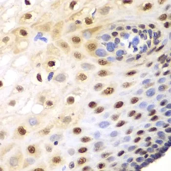
ICC/IF analysis of HeLa cells using GTX31811 SRP1 antibody. Working concentration : 2.5 μg/ml


ICC/IF analysis of HeLa cells using GTX31811 SRP1 antibody. Working concentration : 2.5 μg/ml
SRP1 antibody
GTX31811
ApplicationsImmunoFluorescence, Western Blot, ELISA, ImmunoCytoChemistry
Product group Antibodies
ReactivityHuman, Mouse, Rat
TargetKPNA1
Overview
- SupplierGeneTex
- Product NameSRP1 antibody
- Delivery Days Customer9
- Application Supplier NoteWB: 1 microg/mL. ICC/IF: 2.5 microg/mL. *Optimal dilutions/concentrations should be determined by the researcher.Not tested in other applications.
- ApplicationsImmunoFluorescence, Western Blot, ELISA, ImmunoCytoChemistry
- CertificationResearch Use Only
- ClonalityPolyclonal
- Concentration1 mg/ml
- ConjugateUnconjugated
- Gene ID3836
- Target nameKPNA1
- Target descriptionkaryopherin subunit alpha 1
- Target synonymsIPOA5, NPI-1, RCH2, SRP1, importin subunit alpha-5, RAG cohort protein 2, SRP1-beta, importin alpha 5, importin subunit alpha-1, importin-alpha-S1, karyopherin alpha 1 (importin alpha 5), nucleoprotein interactor 1, recombination activating gene cohort 2
- HostRabbit
- IsotypeIgG
- Protein IDP52294
- Protein NameImportin subunit alpha-5
- Scientific DescriptionThe transport of molecules between the nucleus and the cytoplasm in eukaryotic cells is mediated by the nuclear pore complex (NPC), which consists of 60-100 proteins. Small molecules (up to 70 kD) can pass through the nuclear pore by nonselective diffusion while larger molecules are transported by an active process. The protein encoded by this gene belongs to the importin alpha family, and is involved in nuclear protein import. This protein interacts with the recombination activating gene 1 (RAG1) protein and is a putative substrate of the RAG1 ubiquitin ligase. Alternative splicing results in multiple transcript variants. [provided by RefSeq, Nov 2012]
- ReactivityHuman, Mouse, Rat
- Storage Instruction-20°C or -80°C,2°C to 8°C
- UNSPSC41116161
Datasheet
Related products
Product group Antibodies
KPNA1 AntibodyCSB-PA012483LA01HU
ApplicationsELISA, ImmunoHistoChemistry
ReactivityHuman
TargetKPNA1
- SizePrice
Product group Antibodies

KPNA1 / Importin Alpha 5 AntibodyLS-C748664
ApplicationsImmunoFluorescence, Western Blot, ImmunoHistoChemistry
ReactivityHuman, Mouse, Rat
TargetKPNA1
- SizePrice

Product group Antibodies
Anti-KPNA1 AntibodyHPA053627
ApplicationsWestern Blot, ImmunoCytoChemistry, ImmunoHistoChemistry
ReactivityHuman
TargetKPNA1
- SizePrice
Product group Antibodies

ApplicationsWestern Blot, ELISA
ReactivityBovine, Canine, Human, Mouse, Porcine, Rat
TargetKPNA1
- SizePrice

Product group Antibodies
Anti-SRP1/KPNA1 Antibody Picoband(r)A04024-1-CARRIER-FREE
ApplicationsFlow Cytometry, ImmunoFluorescence, Western Blot, ImmunoCytoChemistry
ReactivityHuman, Mouse, Rat
TargetKPNA1
- SizePrice
Product group Antibodies
SRP1 antibody, N-termGTX26035
ApplicationsWestern Blot
ReactivityHuman
TargetKPNA1
- SizePrice
Product group Antibodies
ApplicationsImmunoPrecipitation, Western Blot, ImmunoCytoChemistry, ImmunoHistoChemistry
ReactivityMouse
TargetKPNA1
- SizePrice
Product group Antibodies
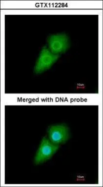
SRP1 antibodyGTX112284
ApplicationsImmunoFluorescence, Western Blot, ImmunoCytoChemistry
ReactivityHuman, Mouse
TargetKPNA1
- SizePrice
Product group Antibodies
SRP1 antibodyGTX54302
ApplicationsImmunoFluorescence, Western Blot, ImmunoCytoChemistry, ImmunoHistoChemistry, ImmunoHistoChemistry Paraffin
ReactivityHuman, Mouse
TargetKPNA1
- SizePrice