

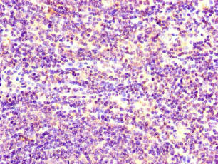
Immunohistochemistry of paraffin-embedded human lymph node tissue using CSB-PA022829LA01HU at dilution of 1:100
Immunohistochemistry of paraffin-embedded human lymph node tissue using CSB-PA022829LA01HU at dilution of 1:100
STIM1 Antibody
CSB-PA022829LA01HU
ApplicationsELISA, ImmunoHistoChemistry
Product group Antibodies
ReactivityHuman
TargetSTIM1
Overview
- SupplierCusabio
- Product NameSTIM1 Antibody
- Delivery Days Customer20
- ApplicationsELISA, ImmunoHistoChemistry
- CertificationResearch Use Only
- ClonalityPolyclonal
- ConjugateUnconjugated
- Gene ID6786
- Target nameSTIM1
- Target descriptionstromal interaction molecule 1
- Target synonymsD11S4896E, GOK, IMD10, STRMK, TAM, TAM1, stromal interaction molecule 1
- HostRabbit
- IsotypeIgG
- Protein IDQ13586
- Protein NameStromal interaction molecule 1
- Scientific DescriptionPlays a role in mediating store-operated Ca(2+) entry (SOCE), a Ca(2+) influx following depletion of intracellular Ca(2+) stores (PubMed:15866891, PubMed:16005298, PubMed:16208375, PubMed:16537481, PubMed:16733527, PubMed:16766533, PubMed:16807233, PubMed:18854159, PubMed:19249086, PubMed:22464749, PubMed:24069340, PubMed:24351972, PubMed:24591628, PubMed:26322679, PubMed:25326555). Acts as Ca(2+) sensor in the endoplasmic reticulum via its EF-hand domain. Upon Ca(2+) depletion, translocates from the endoplasmic reticulum to the plasma membrane where it activates the Ca(2+) release-activated Ca(2+) (CRAC) channel subunit ORAI1 (PubMed:16208375, PubMed:16537481). Involved in enamel formation (PubMed:24621671). Activated following interaction with TMEM110/STIMATE, leading to promote STIM1 conformational switch (PubMed:26322679).
- ReactivityHuman
- Storage Instruction-20°C or -80°C
- UNSPSC41116161
Related products
Product group Antibodies
ApplicationsFlow Cytometry, Western Blot, ELISA
ReactivityHuman
- SizePrice
Product group Antibodies
Anti-STIM1 Antibody144-66387
ApplicationsImmunoFluorescence, Western Blot
ReactivityHuman, Mouse, Rat
TargetSTIM1
- SizePrice
Product group Antibodies

GOK / STIM1 Antibody (Biotin)LS-C682257
ApplicationsELISA
ReactivityHuman
TargetSTIM1
- SizePrice

Product group Antibodies
Anti-STIM1 Antibody Picoband(r)A00312-1-CARRIER-FREE
ApplicationsFlow Cytometry, ImmunoFluorescence, Western Blot, ELISA, ImmunoHistoChemistry
ReactivityHuman, Mouse, Rat
TargetSTIM1
- SizePrice
Product group Antibodies
STIM1 Recombinant Antibody, Biotin ConjugatedBSM-61325R-BIOTIN
ApplicationsImmunoPrecipitation, Western Blot, ImmunoHistoChemistry, ImmunoHistoChemistry Frozen, ImmunoHistoChemistry Paraffin
TargetSTIM1
- SizePrice
Product group Antibodies
References

Goat anti-STIM1EB07469
ApplicationsFlow Cytometry, Western Blot, ELISA
ReactivityBovine, Canine, Human, Mouse, Rat
TargetSTIM1
- SizePrice
Product group Antibodies
ApplicationsImmunoPrecipitation, Western Blot, ImmunoCytoChemistry, ImmunoHistoChemistry
ReactivityMouse, Rat
TargetSTIM1
- SizePrice

Product group Antibodies
Anti-STIM1 AntibodyHPA011088
ApplicationsWestern Blot, ImmunoCytoChemistry, ImmunoHistoChemistry
ReactivityHuman
TargetSTIM1
- SizePrice
Product group Antibodies
Anti-STIM1 AntibodyCAB7411
ApplicationsImmunoFluorescence, ImmunoPrecipitation, Western Blot, ELISA, ImmunoCytoChemistry
ReactivityHuman
TargetSTIM1
- SizePrice
Product group Antibodies
TargetSTIM1
- SizePrice