

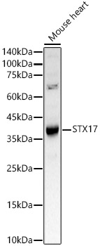
Various tissue extracts (50 μg) were separated by 12% SDS-PAGE, and the membrane was blotted with STX17 antibody [HL3534] (GTX641431) diluted at 1:1000. The HRP-conjugated anti-rabbit IgG antibody (GTX213110-01) was used to detect the primary antibody.
![Wild-type (WT) and STX17 knockout (KO) HeLa cell extracts (5 μg) were separated by 12% SDS-PAGE, and the membrane was blotted with STX17 antibody [HL3534] (GTX641431) diluted at 1:1000. The HRP-conjugated anti-rabbit IgG antibody (GTX213110-01) was used to detect the primary antibody, and the signal was developed with Trident ECL plus-Enhanced. Corresponding RNA expression data for the same cell lines are based on Human Protein Atlas program.](https://www.genetex.com/upload/website/prouct_img/normal/GTX641431/GTX641431_T-45614_20241220_WB_KO_watermark_24122400_460.webp "Wild-type (WT) and STX17 knockout (KO) HeLa cell extracts (5 μg) were separated by 12% SDS-PAGE, and the membrane was blotted with STX17 antibody [HL3534] (GTX641431) diluted at 1:1000. The HRP-conjugated anti-rabbit IgG antibody (GTX213110-01) was used to detect the primary antibody, and the signal was developed with Trident ECL plus-Enhanced. Corresponding RNA expression data for the same cell lines are based on Human Protein Atlas program.")
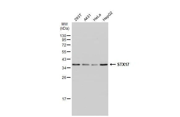
![Various whole cell extracts (30 μg) were separated by 12% SDS-PAGE, and the membrane was blotted with STX17 antibody [HL3534] (GTX641431) diluted at 1:1000. The HRP-conjugated anti-rabbit IgG antibody (GTX213110-01) was used to detect the primary antibody.](https://www.genetex.com/upload/website/prouct_img/normal/GTX641431/GTX641431_T-45614_20241220_WB_24122400_753.webp "Various whole cell extracts (30 μg) were separated by 12% SDS-PAGE, and the membrane was blotted with STX17 antibody [HL3534] (GTX641431) diluted at 1:1000. The HRP-conjugated anti-rabbit IgG antibody (GTX213110-01) was used to detect the primary antibody.")
![STX17 antibody [HL3534] detects STX17 protein by immunohistochemical analysis. Sample: Paraffin-embedded mouse testis. STX17 stained by STX17 antibody [HL3534] (GTX641431) diluted at 1:200. Antigen Retrieval: Citrate buffer, pH 6.0, 15 min](https://www.genetex.com/upload/website/prouct_img/normal/GTX641431/GTX641431_T-45614_20250128_IHC-P_M_25021923_401.webp "STX17 antibody [HL3534] detects STX17 protein by immunohistochemical analysis. Sample: Paraffin-embedded mouse testis. STX17 stained by STX17 antibody [HL3534] (GTX641431) diluted at 1:200. Antigen Retrieval: Citrate buffer, pH 6.0, 15 min")
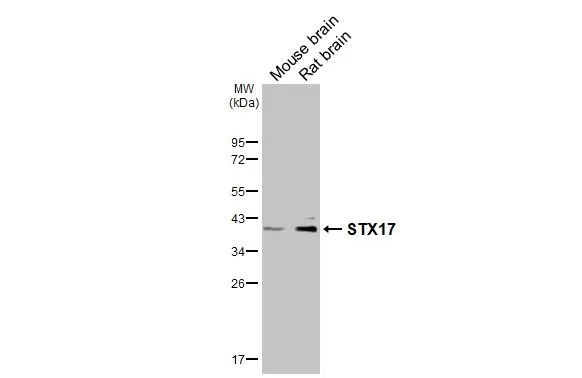
Various tissue extracts (50 μg) were separated by 12% SDS-PAGE, and the membrane was blotted with STX17 antibody [HL3534] (GTX641431) diluted at 1:1000. The HRP-conjugated anti-rabbit IgG antibody (GTX213110-01) was used to detect the primary antibody.
STX17 antibody [HL3534]
GTX641431
ApplicationsWestern Blot, ImmunoHistoChemistry, ImmunoHistoChemistry Paraffin
Product group Antibodies
ReactivityHuman, Mouse, Rat
TargetSTX17
Overview
- SupplierGeneTex
- Product NameSTX17 antibody [HL3534]
- Delivery Days Customer9
- Application Supplier NoteWB: 1:500-1:3000. *Optimal dilutions/concentrations should be determined by the researcher.Not tested in other applications.
- ApplicationsWestern Blot, ImmunoHistoChemistry, ImmunoHistoChemistry Paraffin
- CertificationResearch Use Only
- ClonalityMonoclonal
- Clone IDHL3534
- Concentration1 mg/ml
- ConjugateUnconjugated
- Gene ID55014
- Target nameSTX17
- Target descriptionsyntaxin 17
- Target synonymssyntaxin-17
- HostRabbit
- IsotypeIgG
- Protein IDP56962
- Protein NameSyntaxin-17
- Scientific DescriptionEnables SNAP receptor activity; SNARE binding activity; and protein phosphatase binding activity. Involved in several processes, including autophagosome membrane docking; endoplasmic reticulum to Golgi vesicle-mediated transport; and endoplasmic reticulum-Golgi intermediate compartment organization. Acts upstream of or within protein localization to phagophore assembly site. Located in several cellular components, including autophagosome membrane; endoplasmic reticulum-Golgi intermediate compartment; and mitochondria-associated endoplasmic reticulum membrane contact site. Part of SNARE complex. [provided by Alliance of Genome Resources, Dec 2024]
- ReactivityHuman, Mouse, Rat
- Storage Instruction-20°C or -80°C,2°C to 8°C
- UNSPSC41116161
Related products
Product group Antibodies
Anti-STX17 AntibodyA93329
ApplicationsImmunoFluorescence, Western Blot, ImmunoCytoChemistry
ReactivityHuman, Mouse
- SizePrice
Product group Antibodies
STX17 AntibodyCSB-PA022892GA01HU
ApplicationsWestern Blot, ELISA, ImmunoHistoChemistry
ReactivityHuman, Mouse, Rat
TargetSTX17
- SizePrice

Product group Antibodies
Anti-STX17 AntibodyHPA001204
ApplicationsWestern Blot, ImmunoHistoChemistry
ReactivityHuman
TargetSTX17
- SizePrice
Product group Antibodies
STX17 antibodyGTX130212
ApplicationsImmunoFluorescence, ImmunoPrecipitation, Western Blot, ImmunoCytoChemistry, ImmunoHistoChemistry, ImmunoHistoChemistry Paraffin
ReactivityHuman, Mouse, Rat
TargetSTX17
- SizePrice
Product group Antibodies

STX17 / Syntaxin 17 Antibody (aa100-150)LS-C763256
ApplicationsImmunoPrecipitation
ReactivityHuman
TargetSTX17
- SizePrice