

![SUMO-2/3(SM23/496), Biotin conjugate, 0.1mg/mL [26628-22-8]](https://biotium.com/wp-content/uploads/2016/12/BNUB0496-1-1-e1619215712855.jpg "SUMO-2/3(SM23/496), Biotin conjugate, 0.1mg/mL [26628-22-8]")
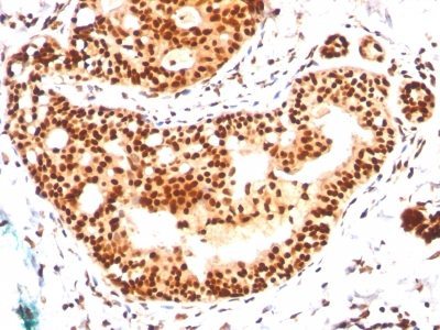
SUMO-2/3(SM23/496), Biotin conjugate, 0.1mg/mL [26628-22-8]
BNCB0496
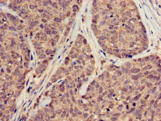
ApplicationsFlow Cytometry, ImmunoFluorescence, Western Blot, ImmunoHistoChemistry, ImmunoHistoChemistry Paraffin
Product group Antibodies
ReactivityBovine, Human, Mouse
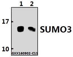
TargetSUMO3
Overview
- SupplierBiotium
- Product NameSUMO-2/3(SM23/496), Biotin conjugate, 0.1mg/mL [26628-22-8]
- Delivery Days Customer9
- ApplicationsFlow Cytometry, ImmunoFluorescence, Western Blot, ImmunoHistoChemistry, ImmunoHistoChemistry Paraffin
- CAS Number26628-22-8
- CertificationResearch Use Only
- ClonalityMonoclonal
- Clone IDSM23/496
- Concentration0.1 mg/ml
- ConjugateBiotin
- Gene ID6612
- Target nameSUMO3
- Target descriptionsmall ubiquitin like modifier 3
- Target synonymsSMT3A, SMT3H1, SUMO-3, Smt3B, small ubiquitin-related modifier 3, SMT3 suppressor of mif two 3 homolog 1, SMT3 suppressor of mif two 3 homolog 3, ubiquitin-like protein SMT3B
- HostMouse
- IsotypeIgG1
- Protein IDP55854
- Protein NameSmall ubiquitin-related modifier 3
- Scientific DescriptionThis mAb reacts with both SUMO-2 and SUMO-3. The small ubiquitin-related modifier (SUMO) proteins, which include SUMO-1, 2 and 3, belong to the ubiquitin-like protein family. Like ubiquitin, the SUMO proteins are synthesized as precursor proteins that undergo processing before conjugation to target proteins. Also, both utilize the E1, E2 and E3 cascade enzymes for conjugation. However, SUMO and ubiquitin differ with respect to targeting. Ubiquitination predominantly targets proteins for degradation, whereas sumoylation targets proteins to a variety of cellular processing, including nuclear transport, transcriptional regulation, apoptosis and protein stability. The unconjugated SUMO-1, 2 and 3 proteins localize to the nuclear membrane, nuclear bodies and cytoplasm, respectively. SUMO-1 utilizes Ubc9 for conjugation to several target proteins, which include MDM2, p53, PML and RanGap1. SUMO-2 and 3 contribute to a greater percentage of protein modification than does SUMO-1 and unlike SUMO-1, they can form polymeric chains. In addition, SUMO-3 regulates beta-Amyloid generation and may be critical in the onset or progression of Alzheimers disease. Note: Conjugates of blue fluorescent dyes like CF®405S and CF®405M are not recommended for detecting low abundance targets, because blue dyes have lower fluorescence and can give higher non-specific background than other dye colors.
- SourceAnimal
- ReactivityBovine, Human, Mouse
- Storage Instruction2°C to 8°C,RT
- UNSPSC41116161
MSDS
Related products
Product group Antibodies
Anti-SUMO3 AntibodyA28667
ApplicationsWestern Blot
ReactivityHuman, Mouse, Rat
- SizePrice

Product group Antibodies
Anti-SUMO2/3 Antibody Picoband(r)A01282-2-CARRIER-FREE
ApplicationsFlow Cytometry, ImmunoFluorescence, Western Blot, ELISA, ImmunoCytoChemistry, ImmunoHistoChemistry
ReactivityHuman, Mouse, Rat
TargetSUMO3
- SizePrice
Product group Antibodies
Anti-SUMO3 Antibody144-61168
ApplicationsWestern Blot
ReactivityHuman, Mouse, Rat
TargetSUMO3
- SizePrice

Product group Antibodies

SUMO3 AntibodyLS-C679695
ApplicationsImmunoFluorescence, ELISA, ImmunoHistoChemistry, ImmunoHistoChemistry Paraffin
ReactivityHuman
TargetSUMO3
- SizePrice
Product group Antibodies
Sumo2/3 Recombinant Antibody, AbBy Fluor-350 ConjugatedBSM-61351R-BF350
ApplicationsFlow Cytometry, ImmunoFluorescence
ReactivityHuman, Mouse, Rat
TargetSUMO3
- SizePrice
Product group Antibodies
SUMO3 AntibodyCSB-PA05509A0RB
ApplicationsImmunoFluorescence, ELISA, ImmunoHistoChemistry
ReactivityHuman
TargetSUMO3
- SizePrice
![WB analysis of HeLa whole cell lysate using GTX00699 SUMO2 + SUMO3 antibody [3H12]. High molecular multiple bands were observed.](https://www.genetex.com/upload/website/prouct_img/normal/GTX00699/GTX00699_20191104_WB_w_23053121_569.webp)
Product group Antibodies
SUMO2 + SUMO3 antibody [3H12]GTX00699
ApplicationsImmunoFluorescence, Western Blot, ELISA, ImmunoCytoChemistry, ImmunoHistoChemistry, ImmunoHistoChemistry Frozen
ReactivityHamster, Human, Mouse, Primate, Rat
TargetSUMO3
- SizePrice