

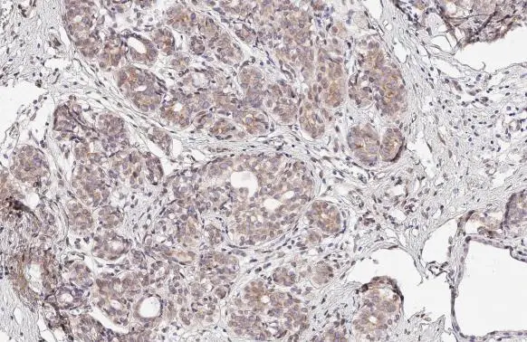
SUN1 antibody [HL1947] detects SUN1 protein at nucleus by immunohistochemical analysis. Sample: Paraffin-embedded human breast carcinoma. SUN1 stained by SUN1 antibody [HL1947] (GTX637784) diluted at 1:100. Antigen Retrieval: Citrate buffer, pH 6.0, 15 min
![Various whole cell extracts (30 μg) were separated by 7.5% SDS-PAGE, and the membrane was blotted with SUN1 antibody [HL1947] (GTX637784) diluted at 1:1000. The HRP-conjugated anti-rabbit IgG antibody (GTX213110-01) was used to detect the primary antibody.](https://www.genetex.com/upload/website/prouct_img/normal/GTX637784/GTX637784_44893_20221223_WB_M_Differentiated_22122722_315.webp "Various whole cell extracts (30 μg) were separated by 7.5% SDS-PAGE, and the membrane was blotted with SUN1 antibody [HL1947] (GTX637784) diluted at 1:1000. The HRP-conjugated anti-rabbit IgG antibody (GTX213110-01) was used to detect the primary antibody.")
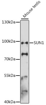
![Various whole cell extracts (50 μg) were separated by 7.5% SDS-PAGE, and the membrane was blotted with SUN1 antibody [HL1947] (GTX637784) diluted at 1:1000. The HRP-conjugated anti-rabbit IgG antibody (GTX213110-01) was used to detect the primary antibody, and the signal was developed with Trident ECL plus-Enhanced.](https://www.genetex.com/upload/website/prouct_img/normal/GTX637784/GTX637784_T-44844_20221223_WB_M_22122722_457.webp "Various whole cell extracts (50 μg) were separated by 7.5% SDS-PAGE, and the membrane was blotted with SUN1 antibody [HL1947] (GTX637784) diluted at 1:1000. The HRP-conjugated anti-rabbit IgG antibody (GTX213110-01) was used to detect the primary antibody, and the signal was developed with Trident ECL plus-Enhanced.")
![SUN1 antibody [HL1947] detects SUN1 protein at nuclear envelope by immunofluorescent analysis. Sample: A431 cells were fixed in 4% paraformaldehyde at RT for 15 min. Green: SUN1 stained by SUN1 antibody [HL1947] (GTX637784) diluted at 1:500. Red: alpha Tubulin, a cytoskeleton marker, stained by alpha Tubulin antibody [GT114] (GTX628802) diluted at 1:1000. Blue: Fluoroshield with DAPI (GTX30920).](https://www.genetex.com/upload/website/prouct_img/normal/GTX637784/GTX637784_T-44844_20230106_ICC_IF_23013122_304.webp "SUN1 antibody [HL1947] detects SUN1 protein at nuclear envelope by immunofluorescent analysis. Sample: A431 cells were fixed in 4% paraformaldehyde at RT for 15 min. Green: SUN1 stained by SUN1 antibody [HL1947] (GTX637784) diluted at 1:500. Red: alpha Tubulin, a cytoskeleton marker, stained by alpha Tubulin antibody [GT114] (GTX628802) diluted at 1:1000. Blue: Fluoroshield with DAPI (GTX30920).")
![Non-transfected (–) and transfected (+) HeLa whole cell extracts (30 μg) were separated by 7.5% SDS-PAGE, and the membrane was blotted with SUN1 antibody [HL1947] (GTX637784) diluted at 1:1000. The HRP-conjugated anti-rabbit IgG antibody (GTX213110-01) was used to detect the primary antibody, and the signal was developed with Trident ECL plus-Enhanced.](https://www.genetex.com/upload/website/prouct_img/normal/GTX637784/GTX637784_44893_20230217_WB_shRNA_watermark_23022022_151.webp "Non-transfected (–) and transfected (+) HeLa whole cell extracts (30 μg) were separated by 7.5% SDS-PAGE, and the membrane was blotted with SUN1 antibody [HL1947] (GTX637784) diluted at 1:1000. The HRP-conjugated anti-rabbit IgG antibody (GTX213110-01) was used to detect the primary antibody, and the signal was developed with Trident ECL plus-Enhanced.")
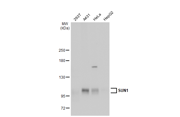
![Various whole cell extracts (30 μg) were separated by 5% SDS-PAGE, and the membrane was blotted with SUN1 antibody [HL1947] (GTX637784) diluted at 1:1000. The HRP-conjugated anti-rabbit IgG antibody (GTX213110-01) was used to detect the primary antibody. Corresponding RNA expression data for the same cell lines are based on Human Protein Atlas program.](https://www.genetex.com/upload/website/prouct_img/normal/GTX637784/GTX637784_44893_20230505_WB_TPM_watermark_23050918_859.webp "Various whole cell extracts (30 μg) were separated by 5% SDS-PAGE, and the membrane was blotted with SUN1 antibody [HL1947] (GTX637784) diluted at 1:1000. The HRP-conjugated anti-rabbit IgG antibody (GTX213110-01) was used to detect the primary antibody. Corresponding RNA expression data for the same cell lines are based on Human Protein Atlas program.")
![Various whole cell extracts (30 μg) were separated by 5% SDS-PAGE, and the membrane was blotted with SUN1 antibody [HL1947] (GTX637784) diluted at 1:1000. The HRP-conjugated anti-rabbit IgG antibody (GTX213110-01) was used to detect the primary antibody. Corresponding RNA expression data for the same cell lines are based on Human Protein Atlas program.](https://www.genetex.com/upload/website/prouct_img/normal/GTX637784/GTX637784_45278_20240112_WB_TPM_watermark_24122400_528.webp "Various whole cell extracts (30 μg) were separated by 5% SDS-PAGE, and the membrane was blotted with SUN1 antibody [HL1947] (GTX637784) diluted at 1:1000. The HRP-conjugated anti-rabbit IgG antibody (GTX213110-01) was used to detect the primary antibody. Corresponding RNA expression data for the same cell lines are based on Human Protein Atlas program.")
SUN1 antibody [HL1947] detects SUN1 protein at nucleus by immunohistochemical analysis. Sample: Paraffin-embedded human breast carcinoma. SUN1 stained by SUN1 antibody [HL1947] (GTX637784) diluted at 1:100. Antigen Retrieval: Citrate buffer, pH 6.0, 15 min
SUN1 antibody [HL1947]
GTX637784
ApplicationsImmunoFluorescence, Western Blot, ImmunoCytoChemistry, ImmunoHistoChemistry, ImmunoHistoChemistry Paraffin
Product group Antibodies
ReactivityHuman, Mouse
TargetSUN1
Overview
- SupplierGeneTex
- Product NameSUN1 antibody [HL1947]
- Delivery Days Customer9
- Application Supplier NoteWB: 1:500-1:3000. *Optimal dilutions/concentrations should be determined by the researcher.Not tested in other applications.
- ApplicationsImmunoFluorescence, Western Blot, ImmunoCytoChemistry, ImmunoHistoChemistry, ImmunoHistoChemistry Paraffin
- CertificationResearch Use Only
- ClonalityMonoclonal
- Clone IDHL1947
- Concentration1 mg/ml
- ConjugateUnconjugated
- Gene ID23353
- Target nameSUN1
- Target descriptionSad1 and UNC84 domain containing 1
- Target synonymsUNC84A, SUN domain-containing protein 1, Sad1 unc-84 domain protein 1, protein unc-84 homolog A, sad1/unc-84 protein-like 1, unc-84 homolog A
- HostRabbit
- IsotypeIgG
- Protein IDO94901
- Protein NameSUN domain-containing protein 1
- Scientific DescriptionThis gene is a member of the unc-84 homolog family and encodes a nuclear nuclear envelope protein with an Unc84 (SUN) domain. The protein is involved in nuclear anchorage and migration. Alternatively spliced transcript variants have been described. [provided by RefSeq, Jan 2010]
- ReactivityHuman, Mouse
- Storage Instruction-20°C or -80°C,2°C to 8°C
- UNSPSC41116161
Datasheet
Related products
Product group Antibodies
Anti-SUN1 AntibodyA91425
ApplicationsWestern Blot
ReactivityHuman, Mouse, Rat
- SizePrice
Product group Antibodies
SUN1 Recombinant AntibodyBSM-54420R
ApplicationsFlow Cytometry, ImmunoFluorescence, Western Blot, ImmunoCytoChemistry, ImmunoHistoChemistry, ImmunoHistoChemistry Paraffin
ReactivityHuman, Rat
TargetSUN1
- SizePrice
Product group Antibodies
Sun1 Polyclonal AntibodyCAC11632
ApplicationsWestern Blot, ELISA
ReactivityPlant
- SizePrice
Product group Antibodies
SUN1 AntibodyCSB-PA24689A0RB
ApplicationsWestern Blot, ELISA
ReactivityPlant
TargetSUN1
- SizePrice

Product group Antibodies
Anti-SUN1 AntibodyHPA008461
ApplicationsWestern Blot, ImmunoCytoChemistry, ImmunoHistoChemistry
ReactivityHuman
TargetSUN1
- SizePrice
Product group Antibodies
SUN1 antibodyGTX115697
ApplicationsWestern Blot
ReactivityHuman
TargetSUN1
- SizePrice
Product group Antibodies
References
SUN1 antibody, N-termGTX45959
ApplicationsWestern Blot, ImmunoHistoChemistry, ImmunoHistoChemistry Paraffin
ReactivityCanine, Human, Mouse
TargetSUN1
- SizePrice
![SUN1 antibody [HL1946] detects SUN1 protein at nuclear envelope by immunofluorescent analysis. Sample: HeLa cells were fixed in 4% paraformaldehyde at RT for 15 min. Green: SUN1 stained by SUN1 antibody [HL1946] (GTX637783) diluted at 1:500. Blue: Fluoroshield with DAPI (GTX30920).](https://www.genetex.com/upload/website/prouct_img/normal/GTX637783/GTX637783_T-44844_20221104_ICC_IF_22112219_191.webp)
Product group Antibodies
SUN1 antibody [HL1946]GTX637783
ApplicationsImmunoFluorescence, ImmunoCytoChemistry
ReactivityHuman
TargetSUN1
- SizePrice
Product group Antibodies

SUN1 AntibodyLS-C752215
ApplicationsWestern Blot, ELISA
ReactivityHuman
TargetSUN1
- SizePrice