

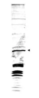
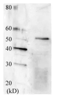
Western blot analysis is shown using GeneTex's Affinity Purified anti-Swi6 antibody to detect endogenous protein present in S.pombe lysate (arrowhead). Comparison to a molecular weight marker (not shown) indicates a band of ~43 kDa corresponding to S.pombe Swi6 protein. ~35ug of lysate was loaded per lane onto a 4-20% gradient gel for SDS-PAGE followed by transfer to 0.45 mm nitrocellulose. The blot was incubated with a 1:1,700 dilution of the antibody at room temperature for 2 h followed by detection using IRDye?800 labeled Goat-a-Rabbit IgG [H&L] diluted 1:5,000 for 45 min. IRDye?800 fluorescence image was captured using the OdysseyR Infrared Imaging System developed by LI-COR. IRDye is a trademark of LI-COR, Inc. Other detection systems will yield similar results.

Western blot analysis is shown using GeneTex's Affinity Purified anti-Swi6 antibody to detect endogenous protein present in S.pombe lysate (arrowhead). Comparison to a molecular weight marker (not shown) indicates a band of ~43 kDa corresponding to S.pombe Swi6 protein. ~35ug of lysate was loaded per lane onto a 4-20% gradient gel for SDS-PAGE followed by transfer to 0.45 mm nitrocellulose. The blot was incubated with a 1:1,700 dilution of the antibody at room temperature for 2 h followed by detection using IRDye?800 labeled Goat-a-Rabbit IgG [H&L] diluted 1:5,000 for 45 min. IRDye?800 fluorescence image was captured using the OdysseyR Infrared Imaging System developed by LI-COR. IRDye is a trademark of LI-COR, Inc. Other detection systems will yield similar results.
Swi6 antibody
GTX48740
ApplicationsWestern Blot, ELISA
Product group Antibodies
ReactivityYeast
Targetswi6
Overview
- SupplierGeneTex
- Product NameSwi6 antibody
- Delivery Days Customer9
- Application Supplier NoteWB: 1:500-1:3000. ELISA: 1:5000-1:30000. *Optimal dilutions/concentrations should be determined by the researcher.Not tested in other applications.
- ApplicationsWestern Blot, ELISA
- CertificationResearch Use Only
- ClonalityPolyclonal
- Concentration1 mg/ml
- ConjugateUnconjugated
- Gene ID2541633
- Target nameswi6
- Target descriptionheterochromatin (HP1) family chromodomain protein Swi6
- Target synonymsSPOM_SPAC664.01C, SPAC824.10c, heterochromatin (HP1) family chromodomain protein Swi6
- HostRabbit
- IsotypeIgG
- Protein IDP40381
- Protein NameChromatin-associated protein swi6
- ReactivityYeast
- Storage Instruction-20°C or -80°C,2°C to 8°C
- UNSPSC41116161
Datasheet
Related products
Product group Antibodies
swi6 AntibodyCSB-PA340241XA01SXV
ApplicationsWestern Blot, ELISA
ReactivityYeast
Targetswi6
- SizePrice
Product group Antibodies
Swi6 (S. pombe) antibodyGTX64174
ApplicationsImmunoPrecipitation, Western Blot, ChIP Chromatin ImmunoPrecipitation
ReactivityYeast
Targetswi6
- SizePrice