

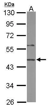
Sample (50 ug of whole cell lysate) A: mouse liver 10% SDS PAGE GTX120025 diluted at 1:500
![SYAP1 antibody [N3C3] detects SYAP1 protein at nucleus and Golgi apparatus by immunofluorescent analysis. Sample: A375 cells were fixed in 4% paraformaldehyde at RT for 15 min. Green: SYAP1 protein stained by SYAP1 antibody [N3C3] (GTX120025) diluted at 1:500. Blue: Hoechst 33342 staining. Scale bar = 10 μm.](https://www.genetex.com/upload/website/prouct_img/normal/GTX120025/GTX120025_40373_IFA_w_23060519_869.webp "SYAP1 antibody [N3C3] detects SYAP1 protein at nucleus and Golgi apparatus by immunofluorescent analysis. Sample: A375 cells were fixed in 4% paraformaldehyde at RT for 15 min. Green: SYAP1 protein stained by SYAP1 antibody [N3C3] (GTX120025) diluted at 1:500. Blue: Hoechst 33342 staining. Scale bar = 10 μm.")
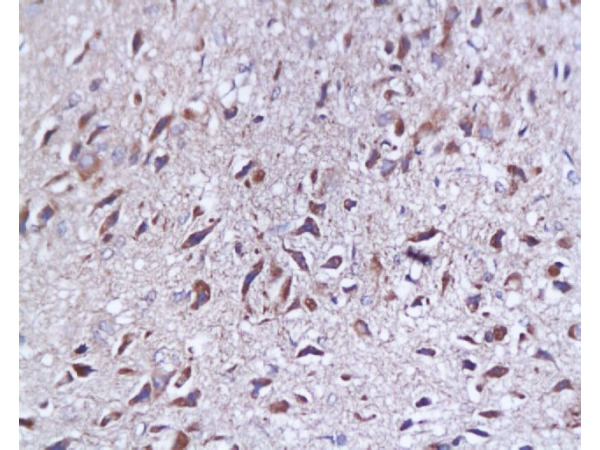
 dilution: 1:500.
Antigen Retrieval: Trilogy? (EDTA based, pH 8.0) buffer, 15min")
 A: 293T 10% SDS PAGE GTX120025 diluted at 1:1000")
Sample (50 ug of whole cell lysate) A: mouse liver 10% SDS PAGE GTX120025 diluted at 1:500
SYAP1 antibody [N3C3]
GTX120025
ApplicationsImmunoFluorescence, Western Blot, ImmunoCytoChemistry, ImmunoHistoChemistry, ImmunoHistoChemistry Paraffin
Product group Antibodies
ReactivityHuman, Mouse
TargetSYAP1
Overview
- SupplierGeneTex
- Product NameSYAP1 antibody [N3C3]
- Delivery Days Customer9
- Application Supplier NoteWB: 1:500-1:3000. ICC/IF: 1:100-1:1000. IHC-P: 1:100-1:1000. *Optimal dilutions/concentrations should be determined by the researcher.Not tested in other applications.
- ApplicationsImmunoFluorescence, Western Blot, ImmunoCytoChemistry, ImmunoHistoChemistry, ImmunoHistoChemistry Paraffin
- CertificationResearch Use Only
- ClonalityPolyclonal
- Concentration1 mg/ml
- ConjugateUnconjugated
- Gene ID94056
- Target nameSYAP1
- Target descriptionsynapse associated protein 1
- Target synonymsBSTA, PRO3113, synapse-associated protein 1, BSD domain-containing signal transducer and Akt interactor protein, SAP47 homolog, synapse associated protein 1, SAP47 homolog
- HostRabbit
- IsotypeIgG
- Protein IDQ96A49
- Protein NameSynapse-associated protein 1
- ReactivityHuman, Mouse
- Storage Instruction-20°C or -80°C,2°C to 8°C
- UNSPSC41116161
Datasheet
Related products
Product group Antibodies

SYAP1 Antibody (aa25-75)LS-C762410
ApplicationsImmunoPrecipitation
ReactivityHuman
TargetSYAP1
- SizePrice
Product group Antibodies
SYAP1 Polyclonal AntibodyBS-0121R
ApplicationsFlow Cytometry, ImmunoFluorescence, Western Blot, ELISA, ImmunoCytoChemistry, ImmunoHistoChemistry, ImmunoHistoChemistry Frozen, ImmunoHistoChemistry Paraffin
ReactivityBovine, Canine, Chicken, Equine, Human, Mouse, Porcine, Rat
TargetSYAP1
- SizePrice
Product group Antibodies
SYAP1 AntibodyCSB-PA846581XA01HU
ApplicationsWestern Blot, ELISA
ReactivityHuman
TargetSYAP1
- SizePrice

Product group Antibodies
Anti-SYAP1 AntibodyHPA000175
ApplicationsImmunoHistoChemistry
ReactivityHuman
TargetSYAP1
- SizePrice