

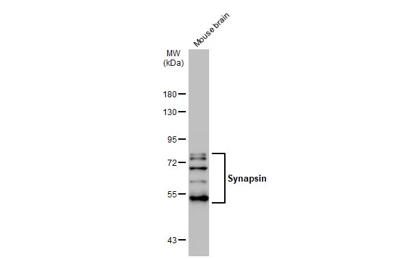
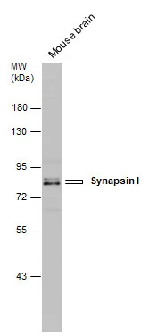
Mouse tissue extract (50 μg) was separated by 7.5% SDS-PAGE, and the membrane was blotted with Synapsin antibody (GTX109960) diluted at 1:1000. The HRP-conjugated anti-rabbit IgG antibody (GTX213110-01) was used to detect the primary antibody.
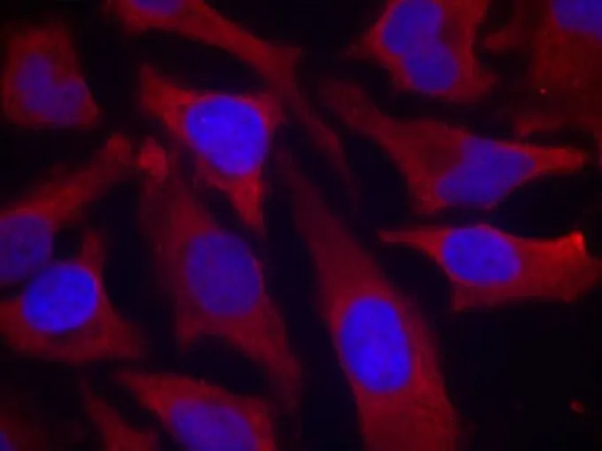
![Synapsin antibody detects Synapsin protein by immunofluorescent analysis. Sample: DIV10 rat E18 primary cortical neuron cells were fixed in 4% paraformaldehyde at RT for 15 min. Green: Synapsin stained by Synapsin antibody (GTX109960) diluted at 1:500. Red: Tau, stained by Tau antibody [GT287] (GTX634809) diluted at 1:500. Blue: Fluoroshield with DAPI (GTX30920).](https://www.genetex.com/upload/website/prouct_img/normal/GTX109960/GTX109960_43628_20191125_ICC_IF_R_w_23060500_753.webp "Synapsin antibody detects Synapsin protein by immunofluorescent analysis. Sample: DIV10 rat E18 primary cortical neuron cells were fixed in 4% paraformaldehyde at RT for 15 min. Green: Synapsin stained by Synapsin antibody (GTX109960) diluted at 1:500. Red: Tau, stained by Tau antibody [GT287] (GTX634809) diluted at 1:500. Blue: Fluoroshield with DAPI (GTX30920).")
Mouse tissue extract (50 μg) was separated by 7.5% SDS-PAGE, and the membrane was blotted with Synapsin antibody (GTX109960) diluted at 1:1000. The HRP-conjugated anti-rabbit IgG antibody (GTX213110-01) was used to detect the primary antibody.
Synapsin antibody
GTX109960
ApplicationsImmunoFluorescence, Western Blot, ImmunoCytoChemistry
Product group Antibodies
ReactivityHuman, Mouse, Rat
TargetSYN1
Overview
- SupplierGeneTex
- Product NameSynapsin antibody
- Delivery Days Customer9
- Application Supplier NoteWB: 1:1000. *Optimal dilutions/concentrations should be determined by the researcher.Not tested in other applications.
- ApplicationsImmunoFluorescence, Western Blot, ImmunoCytoChemistry
- CertificationResearch Use Only
- ClonalityPolyclonal
- Concentration0.25 mg/ml
- ConjugateUnconjugated
- Gene ID6853
- Target nameSYN1
- Target descriptionsynapsin I
- Target synonymsEPILX, EPILX1, MRX50, SYN1a, SYN1b, SYNI, synapsin-1, brain protein 4.1, synapsin Ib
- HostRabbit
- IsotypeIgG
- Protein IDO14994
- Protein NameSynapsin-3
- ReactivityHuman, Mouse, Rat
- Storage Instruction-20°C or -80°C,2°C to 8°C
- UNSPSC41116161
Datasheet
Related products
Product group Antibodies
Anti-SYN1 AntibodyA43930
ApplicationsWestern Blot
ReactivityHuman, Mouse, Rat
- SizePrice
Product group Antibodies
Anti-SYN1 Antibody144-64907
ApplicationsWestern Blot
ReactivityHuman, Mouse, Rat
TargetSYN1
- SizePrice
Product group Antibodies
References
Synapsin 1 Polyclonal AntibodyBS-3501R
ApplicationsImmunoFluorescence, Western Blot, ELISA, ImmunoCytoChemistry, ImmunoHistoChemistry, ImmunoHistoChemistry Frozen, ImmunoHistoChemistry Paraffin
ReactivityHuman, Mouse, Porcine, Rat
TargetSYN1
- SizePrice
Product group Antibodies
SYN1 AntibodyCSB-PA004200
ApplicationsImmunoFluorescence, Western Blot, ELISA, ImmunoHistoChemistry
ReactivityHuman, Mouse, Rat
TargetSYN1
- SizePrice
Product group Antibodies
SYN1 Polyclonal AntibodyCAC13951
ApplicationsWestern Blot, ELISA, ImmunoHistoChemistry
ReactivityMouse
TargetSYN1
- SizePrice
Product group Antibodies

SYN1 / Synapsin 1 AntibodyLS-C402809
ApplicationsWestern Blot, ELISA
ReactivityHuman, Mouse, Rat
TargetSYN1
- SizePrice

Product group Antibodies
Anti-SYN1 AntibodyHPA000397
ApplicationsWestern Blot, ImmunoHistoChemistry
ReactivityHuman
TargetSYN1
- SizePrice
Product group Antibodies
Synapsin I antibodyGTX12239
ApplicationsImmunoPrecipitation, Western Blot
ReactivityBovine, Human, Mouse, Rat
TargetSYN1
- SizePrice
Product group Antibodies
Synapsin I antibodyGTX131233
ApplicationsWestern Blot
ReactivityHuman, Mouse
TargetSYN1
- SizePrice
Product group Antibodies
ApplicationsImmunoFluorescence, Western Blot, ImmunoCytoChemistry
ReactivityHuman, Mouse
TargetSYN1
- SizePrice