

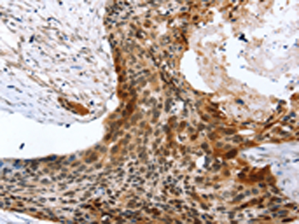
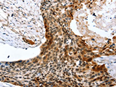
IHC-P analysis of mouse skeletal muscle tissue using GTX85139 SYNPO2 antibody. Working concentration : 20 μg/ml

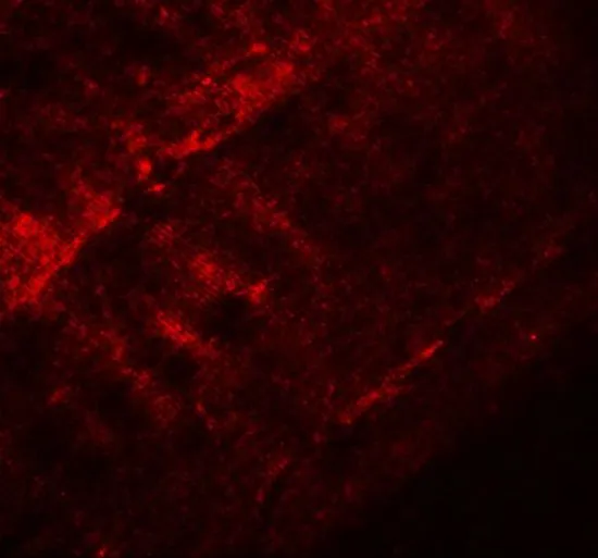
 the absence and (B) the presence of blocking peptide using GTX85139 SYNPO2 antibody. Working concentration : 1 μg/ml")
IHC-P analysis of mouse skeletal muscle tissue using GTX85139 SYNPO2 antibody. Working concentration : 20 μg/ml
SYNPO2 antibody
GTX85139
ApplicationsWestern Blot, ELISA, ImmunoHistoChemistry, ImmunoHistoChemistry Paraffin
Product group Antibodies
ReactivityHuman, Mouse
TargetSYNPO2
Overview
- SupplierGeneTex
- Product NameSYNPO2 antibody
- Delivery Days Customer9
- Application Supplier NoteWB: 1 microg/mL. IHC-P: 5 microg/mL. *Optimal dilutions/concentrations should be determined by the researcher.Not tested in other applications.
- ApplicationsWestern Blot, ELISA, ImmunoHistoChemistry, ImmunoHistoChemistry Paraffin
- CertificationResearch Use Only
- ClonalityPolyclonal
- Concentration1 mg/ml
- ConjugateUnconjugated
- Gene ID171024
- Target nameSYNPO2
- Target descriptionsynaptopodin 2
- Target synonymssynaptopodin-2, genethonin-2, myopodin
- HostRabbit
- IsotypeIgG
- Protein IDQ9UMS6
- Protein NameSynaptopodin-2
- Scientific DescriptionSYNPO2 was initially identified as myopodin, a member of the synaptopodin family that contains one PPXY motif and multiple PXXP motifs. It colocalizes with alpha-actinin and is found at the Z-disc and during stress conditions will translocate to the nucleus, suggesting that it is part of signaling pathways in addition to its function as a structural protein. SYNPO2 has been shown to bind to calmodulin, alpha-actinin, and smooth muscle myosin and will stimulate actin polymerization in a calmodulin dependent manner, consistent with its proposed role in organizing the cytoskeleton. While deletion of SYNPO2 has been reported to be highly correlated with the invasiveness of prostate cancers, other reports suggest that down-regulation of SYNPO2 reduces the invasiveness and motility of prostate cancer cells.
- ReactivityHuman, Mouse
- Storage Instruction-20°C or -80°C,2°C to 8°C
- UNSPSC41116161
References
- Role of chaperone-assisted selective autophagy (CASA) in mechanical stress protection of periodontal ligament cells. Salim C et al., 2022 Jan, J Orofac OrthopRead this paper
Datasheet
Related products

Product group Antibodies
Anti-SYNPO2 Antibody Picoband(r)A07616-1-CARRIER-FREE
ApplicationsFlow Cytometry, Western Blot, ELISA
ReactivityHuman
TargetSYNPO2
- SizePrice
Product group Antibodies
Anti-SYNPO2 AntibodyA45708
ApplicationsImmunoHistoChemistry
ReactivityHuman
- SizePrice

Product group Antibodies
Anti-SYNPO2 AntibodyHPA030665
ApplicationsImmunoHistoChemistry
ReactivityHuman
TargetSYNPO2
- SizePrice
Product group Antibodies
SYNPO2 AntibodyCSB-PA100389
ApplicationsELISA, ImmunoHistoChemistry
ReactivityHuman
TargetSYNPO2
- SizePrice
Product group Antibodies

SYNPO2 / Synaptopodin 2 AntibodyLS-C406860
ApplicationsELISA, ImmunoHistoChemistry
ReactivityHuman
TargetSYNPO2
- SizePrice