

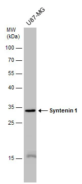
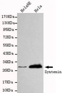
Whole cell extract (30 μg) was separated by 12% SDS-PAGE, and the membrane was blotted with Syntenin 1 antibody [GT1523] (GTX634154) diluted at 1:5000.
![HepG2 whole cell and exosome extracts (1.8 μg) were separated by 12% SDS-PAGE, and the membrane was blotted with Syntenin 1 antibody [GT1523] (GTX634154) diluted at 1:250. The HRP-conjugated anti-mouset IgG antibody (GTX213111-01) was used to detect the primary antibody.](https://www.genetex.com/upload/website/prouct_img/normal/GTX634154/GTX634154_42779_20190115_Fraction_w_23061202_121.webp "HepG2 whole cell and exosome extracts (1.8 μg) were separated by 12% SDS-PAGE, and the membrane was blotted with Syntenin 1 antibody [GT1523] (GTX634154) diluted at 1:250. The HRP-conjugated anti-mouset IgG antibody (GTX213111-01) was used to detect the primary antibody.")
![Rat tissue extract (50 μg) was separated by 12% SDS-PAGE, and the membrane was blotted with Syntenin 1 antibody [GT1523] (GTX634154) diluted at 1:1000. The HRP-conjugated anti-mouse IgG antibody (GTX213111-01) was used to detect the primary antibody, and the signal was developed with Trident ECL plus-Enhanced.](https://www.genetex.com/upload/website/prouct_img/normal/GTX634154/GTX634154_42779_20170922_WB_R_hippocampus_w_23061202_647.webp "Rat tissue extract (50 μg) was separated by 12% SDS-PAGE, and the membrane was blotted with Syntenin 1 antibody [GT1523] (GTX634154) diluted at 1:1000. The HRP-conjugated anti-mouse IgG antibody (GTX213111-01) was used to detect the primary antibody, and the signal was developed with Trident ECL plus-Enhanced.")
![Syntenin 1 antibody [GT1523] detects Syntenin 1 protein by immunofluorescent analysis. Sample: DIV10 rat E18 primary cortical neurons were fixed in 4% paraformaldehyde at RT for 15 min. Green: Syntenin 1 protein stained by Syntenin 1 antibody [GT1523] (GTX634154) diluted at 1:500. Red: beta Tubulin 3/ Tuj1, stained by beta Tubulin 3/ Tuj1 antibody (GTX130245) diluted at 1:500. Blue: Fluoroshield with DAPI (GTX30920).](https://www.genetex.com/upload/website/prouct_img/normal/GTX634154/GTX634154_42779_20170824_IFA_w_23061202_632.webp "Syntenin 1 antibody [GT1523] detects Syntenin 1 protein by immunofluorescent analysis. Sample: DIV10 rat E18 primary cortical neurons were fixed in 4% paraformaldehyde at RT for 15 min. Green: Syntenin 1 protein stained by Syntenin 1 antibody [GT1523] (GTX634154) diluted at 1:500. Red: beta Tubulin 3/ Tuj1, stained by beta Tubulin 3/ Tuj1 antibody (GTX130245) diluted at 1:500. Blue: Fluoroshield with DAPI (GTX30920).")
Whole cell extract (30 μg) was separated by 12% SDS-PAGE, and the membrane was blotted with Syntenin 1 antibody [GT1523] (GTX634154) diluted at 1:5000.
Syntenin 1 antibody [GT1523]
GTX634154
ApplicationsImmunoFluorescence, Western Blot, ImmunoCytoChemistry
Product group Antibodies
ReactivityHuman, Rat
TargetSDCBP
Overview
- SupplierGeneTex
- Product NameSyntenin 1 antibody [GT1523]
- Delivery Days Customer9
- Application Supplier NoteWB: 1:500-1:10000. ICC/IF: 1:100-1:1000. *Optimal dilutions/concentrations should be determined by the researcher.Not tested in other applications.
- ApplicationsImmunoFluorescence, Western Blot, ImmunoCytoChemistry
- CertificationResearch Use Only
- ClonalityMonoclonal
- Clone IDGT1523
- Concentration1 mg/ml
- ConjugateUnconjugated
- Gene ID6386
- Target nameSDCBP
- Target descriptionsyndecan binding protein
- Target synonymsMDA-9, MDA9, SDCBP1, ST1, SYCL, TACIP18, syntenin-1, melanoma differentiation associated protein-9, pro-TGF-alpha cytoplasmic domain-interacting protein 18, scaffold protein Pbp1, syndecan binding protein (syntenin), syndecan-binding protein 1
- HostMouse
- IsotypeIgG2a
- Protein IDO00560
- Protein NameSyntenin-1
- Scientific DescriptionThe protein encoded by this gene was initially identified as a molecule linking syndecan-mediated signaling to the cytoskeleton. The syntenin protein contains tandemly repeated PDZ domains that bind the cytoplasmic, C-terminal domains of a variety of transmembrane proteins. This protein may also affect cytoskeletal-membrane organization, cell adhesion, protein trafficking, and the activation of transcription factors. The protein is primarily localized to membrane-associated adherens junctions and focal adhesions but is also found at the endoplasmic reticulum and nucleus. Alternative splicing results in multiple transcript variants encoding different isoforms. [provided by RefSeq]
- ReactivityHuman, Rat
- Storage Instruction-20°C or -80°C,2°C to 8°C
- UNSPSC12352203
Datasheet
Related products

Product group Antibodies
Anti-Syntenin/SDCBP Antibody Picoband(r)A02475-2-CARRIER-FREE
ApplicationsWestern Blot, ELISA
ReactivityHuman, Mouse, Rat
TargetSDCBP
- SizePrice
Product group Antibodies
Anti-SDCBP Antibody144-05360
ApplicationsImmunoFluorescence, Western Blot
ReactivityHuman, Mouse
TargetSDCBP
- SizePrice
Product group Antibodies
SDCBP Polyclonal AntibodyCAC14759
ApplicationsImmunoFluorescence, Western Blot, ELISA, ImmunoHistoChemistry
ReactivityMouse, Rat
TargetSDCBP
- SizePrice
Product group Antibodies
Syntenin 1 Recombinant Antibody, AbBy Fluor-555 ConjugatedBSM-61948R-BF555
ApplicationsFlow Cytometry, ImmunoFluorescence, Western Blot, ImmunoCytoChemistry
ReactivityHuman
TargetSDCBP
- SizePrice
Product group Antibodies
SDCBP AntibodyCSB-PA020893LA01HU
ApplicationsImmunoFluorescence, Western Blot, ELISA, ImmunoHistoChemistry
ReactivityHuman, Mouse, Rat
TargetSDCBP
- SizePrice
Product group Antibodies

Syntenin AntibodyABX430839
ApplicationsWestern Blot, ELISA, ImmunoHistoChemistry
- SizePrice
Product group Antibodies
Syntenin 1 antibody [3D9-G9-H4]GTX49226
ApplicationsWestern Blot
ReactivityHuman
TargetSDCBP
- SizePrice
Product group Antibodies

ApplicationsWestern Blot, ELISA, ImmunoHistoChemistry
ReactivityCanine, Human, Mouse, Rat
TargetSDCBP
- SizePrice