

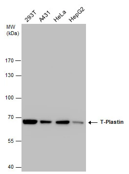
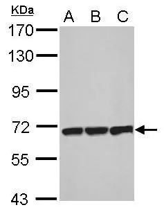
T-Plastin antibody detects T-Plastin protein by western blot analysis. Various whole cell extracts (30 μg) were separated by 7.5% SDS-PAGE, and the membrane was blotted with T-Plastin antibody (GTX632481) diluted at a dilution of 1:1000.
![T-Plastin antibody [GT236] detects T-Plastin protein at cytoplasm by immunohistochemical analysis. Sample: Paraffin-embedded mouse liver. T-Plastin stained by T-Plastin antibody [GT236] (GTX632481) diluted at 1:200. Antigen Retrieval: Citrate buffer, pH 6.0, 15 min](https://www.genetex.com/upload/website/prouct_img/normal/GTX632481/GTX632481_42135_20180622_IHC-P_M_w_23061202_199.webp "T-Plastin antibody [GT236] detects T-Plastin protein at cytoplasm by immunohistochemical analysis. Sample: Paraffin-embedded mouse liver. T-Plastin stained by T-Plastin antibody [GT236] (GTX632481) diluted at 1:200. Antigen Retrieval: Citrate buffer, pH 6.0, 15 min")
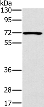
![Non-transfected (–) and transfected (+) 293T whole cell extracts (30 μg) were separated by 7.5% SDS-PAGE, and the membrane was blotted with T-Plastin antibody [GT236] (GTX632481) diluted at 1:1000.](https://www.genetex.com/upload/website/prouct_img/normal/GTX632481/GTX632481_42135_20160825_WB_shRNA_watermark_w_23061202_382.webp "Non-transfected (–) and transfected (+) 293T whole cell extracts (30 μg) were separated by 7.5% SDS-PAGE, and the membrane was blotted with T-Plastin antibody [GT236] (GTX632481) diluted at 1:1000.")
![T-Plastin antibody [GT236] detects T-Plastin protein at cytoplasm by immunohistochemical analysis. Sample: Paraffin-embedded mouse cervix. T-Plastin stained by T-Plastin antibody [GT236] (GTX632481) diluted at 1:200. Antigen Retrieval: Citrate buffer, pH 6.0, 15 min](https://www.genetex.com/upload/website/prouct_img/normal/GTX632481/GTX632481_42135_20180622_IHC-P_M_1_w_23061202_352.webp "T-Plastin antibody [GT236] detects T-Plastin protein at cytoplasm by immunohistochemical analysis. Sample: Paraffin-embedded mouse cervix. T-Plastin stained by T-Plastin antibody [GT236] (GTX632481) diluted at 1:200. Antigen Retrieval: Citrate buffer, pH 6.0, 15 min")
T-Plastin antibody detects T-Plastin protein by western blot analysis. Various whole cell extracts (30 μg) were separated by 7.5% SDS-PAGE, and the membrane was blotted with T-Plastin antibody (GTX632481) diluted at a dilution of 1:1000.
T-Plastin antibody [GT236]
GTX632481
ApplicationsWestern Blot, ImmunoHistoChemistry, ImmunoHistoChemistry Paraffin
Product group Antibodies
ReactivityHuman, Mouse
TargetPLS3
Overview
- SupplierGeneTex
- Product NameT-Plastin antibody [GT236]
- Delivery Days Customer9
- Application Supplier NoteWB: 1:500-1:3000. IHC-P: 1:100-1:1000. *Optimal dilutions/concentrations should be determined by the researcher.Not tested in other applications.
- ApplicationsWestern Blot, ImmunoHistoChemistry, ImmunoHistoChemistry Paraffin
- CertificationResearch Use Only
- ClonalityMonoclonal
- Clone IDGT236
- Concentration1 mg/ml
- ConjugateUnconjugated
- Gene ID5358
- Target namePLS3
- Target descriptionplastin 3
- Target synonymsBMND18, DIH5, T-plastin, plastin-3, T fimbrin, T plastin
- HostMouse
- IsotypeIgG1
- Protein IDP13797
- Protein NamePlastin-3
- Scientific DescriptionPlastins are a family of actin-binding proteins that are conserved throughout eukaryote evolution and expressed in most tissues of higher eukaryotes. In humans, two ubiquitous plastin isoforms (L and T) have been identified. Plastin 1 (otherwise known as Fimbrin) is a third distinct plastin isoform which is specifically expressed at high levels in the small intestine. The L isoform is expressed only in hemopoietic cell lineages, while the T isoform has been found in all other normal cells of solid tissues that have replicative potential (fibroblasts, endothelial cells, epithelial cells, melanocytes, etc.). The C-terminal 570 amino acids of the T-plastin and L-plastin proteins are 83% identical. It contains a potential calcium-binding site near the N terminus. Alternate splicing results in multiple transcript variants.[provided by RefSeq, Feb 2010]
- ReactivityHuman, Mouse
- Storage Instruction-20°C or -80°C,2°C to 8°C
- UNSPSC41116161
Datasheet
Related products
Product group Antibodies
PLS3 AntibodyCSB-PA018206LA01HU
ApplicationsImmunoFluorescence, Western Blot, ELISA, ImmunoHistoChemistry
ReactivityHuman, Mouse
TargetPLS3
- SizePrice
Product group Antibodies
PLS3 Polyclonal AntibodyCAC14863
ApplicationsImmunoFluorescence, Western Blot, ELISA, ImmunoHistoChemistry
ReactivityMouse
TargetPLS3
- SizePrice
Product group Antibodies
Anti-PLS3 Antibody144-03627
ApplicationsImmunoPrecipitation, Western Blot, ImmunoHistoChemistry
ReactivityHuman, Mouse, Rat
TargetPLS3
- SizePrice
Product group Antibodies
Anti-PLS3 AntibodyA38761
ApplicationsWestern Blot, ImmunoHistoChemistry
ReactivityHuman
- SizePrice

Product group Antibodies
Anti-PLS3 AntibodyHPA020433
ApplicationsWestern Blot, ImmunoHistoChemistry
ReactivityHuman
TargetPLS3
- SizePrice

Product group Antibodies
Anti-PLS3 Antibody Picoband(r)A04134-1-CARRIER-FREE
ApplicationsFlow Cytometry, ImmunoFluorescence, ImmunoPrecipitation, Western Blot, ELISA, ImmunoCytoChemistry
ReactivityHuman, Mouse, Rat
TargetPLS3
- SizePrice
Product group Antibodies

PLS3 / T Plastin AntibodyLS-C401936
ApplicationsWestern Blot, ELISA, ImmunoHistoChemistry
ReactivityHuman, Mouse, Rat
TargetPLS3
- SizePrice
Product group Antibodies
PLASTIN3 Polyclonal AntibodyBS-3643R
ApplicationsImmunoFluorescence, Western Blot, ELISA, ImmunoCytoChemistry, ImmunoHistoChemistry, ImmunoHistoChemistry Frozen, ImmunoHistoChemistry Paraffin
ReactivityBovine, Canine, Equine, Human, Mouse, Porcine, Rabbit, Rat
TargetPLS3
- SizePrice
Product group Antibodies
T-Plastin antibodyGTX103323
ApplicationsImmunoFluorescence, Western Blot, ImmunoCytoChemistry, ImmunoHistoChemistry, ImmunoHistoChemistry Paraffin
ReactivityHuman, Mouse
TargetPLS3
- SizePrice