

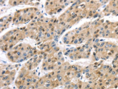
The image on the left is immunohistochemistry of paraffin-embedded Human liver cancer tissue using CSB-PA251266(TAB1 Antibody) at dilution 1/30, on the right is treated with synthetic peptide. (Original magnification: x200)
The image on the left is immunohistochemistry of paraffin-embedded Human liver cancer tissue using CSB-PA251266(TAB1 Antibody) at dilution 1/30, on the right is treated with synthetic peptide. (Original magnification: x200)
TAB1 Antibody
CSB-PA251266
ApplicationsELISA, ImmunoHistoChemistry
Product group Antibodies
ReactivityHuman, Mouse
TargetTAB1
Overview
- SupplierCusabio
- Product NameTAB1 Antibody
- Delivery Days Customer20
- ApplicationsELISA, ImmunoHistoChemistry
- CertificationResearch Use Only
- ClonalityPolyclonal
- ConjugateUnconjugated
- Gene ID10454
- Target nameTAB1
- Target descriptionTGF-beta activated kinase 1 (MAP3K7) binding protein 1
- Target synonyms3'-Tab1, MAP3K7IP1, TGF-beta-activated kinase 1 and MAP3K7-binding protein 1, TAK1-binding protein 1, mitogen-activated protein kinase kinase kinase 7-interacting protein 1, transforming growth factor beta-activated kinase-binding protein 1
- HostRabbit
- IsotypeIgG
- Protein IDQ15750
- Protein NameTGF-beta-activated kinase 1 and MAP3K7-binding protein 1
- Scientific DescriptionThe protein encoded by this gene was identified as a regulator of the MAP kinase kinase kinase MAP3K7/TAK1, which is known to mediate various intracellular signaling pathways, such as those induced by TGF beta, interleukin 1, and WNT-1. This protein interacts and thus activates TAK1 kinase. It has been shown that the C-terminal portion of this protein is sufficient for binding and activation of TAK1, while a portion of the N-terminus acts as a dominant-negative inhibitor of TGF beta, suggesting that this protein may function as a mediator between TGF beta receptors and TAK1. This protein can also interact with and activate the mitogen-activated protein kinase 14 (MAPK14/p38alpha), and thus represents an alternative activation pathway, in addition to the MAPKK pathways, which contributes to the biological responses of MAPK14 to various stimuli. Alternatively spliced transcript variants encoding distinct isoforms have been reported.
- ReactivityHuman, Mouse
- Storage Instruction-20°C or -80°C
- UNSPSC41116161
Related products

Product group Antibodies
Anti-TAB1 Antibody Picoband(r)A02847-1-CARRIER-FREE
ApplicationsWestern Blot, ELISA
ReactivityHuman
TargetTAB1
- SizePrice
Product group Antibodies
Anti-TAB1 Antibody144-05749
ApplicationsImmunoFluorescence, Western Blot
ReactivityHuman, Mouse
TargetTAB1
- SizePrice

Product group Antibodies
Anti-TAB1 AntibodyA14949
ApplicationsImmunoFluorescence, ImmunoPrecipitation, Western Blot, ImmunoCytoChemistry
ReactivityHuman, Mouse
- SizePrice
Product group Antibodies

TAB1 AntibodyLS-C814129
ApplicationsWestern Blot
ReactivityBovine, Canine, Human, Mouse, Porcine, Rat
TargetTAB1
- SizePrice
Product group Antibodies
TAB1 Recombinant AntibodyBSM-62019R
ApplicationsFlow Cytometry, ImmunoFluorescence, ImmunoPrecipitation, Western Blot, ImmunoCytoChemistry, ImmunoHistoChemistry, ImmunoHistoChemistry Frozen, ImmunoHistoChemistry Paraffin
ReactivityHuman, Mouse, Rat
TargetTAB1
- SizePrice
Product group Antibodies
ApplicationsImmunoPrecipitation, Western Blot, ImmunoCytoChemistry, ImmunoHistoChemistry
ReactivityRat
TargetTAB1
- SizePrice
Product group Antibodies
TAB1 antibodyGTX103200
ApplicationsImmunoFluorescence, Western Blot, ImmunoCytoChemistry
ReactivityHuman
TargetTAB1
- SizePrice

Product group Antibodies
Anti-TAB1 AntibodyHPA039988
ApplicationsImmunoCytoChemistry, ImmunoHistoChemistry
ReactivityHuman
TargetTAB1
- SizePrice