

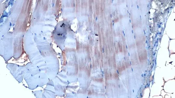
TCEB1 antibody [HL1683] detects TCEB1 protein at cytoplasm by immunohistochemical analysis. Sample: Paraffin-embedded mouse muscle. TCEB1 stained by TCEB1 antibody [HL1683] (GTX637282) diluted at 1:100. Antigen Retrieval: Citrate buffer, pH 6.0, 15 min
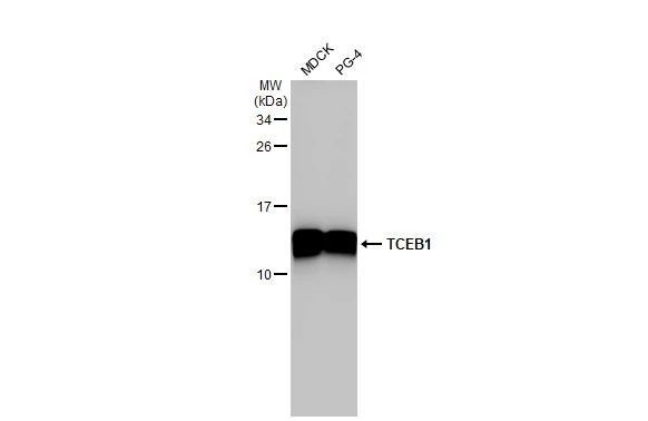
![Various whole cell extracts (30 μg) were separated by 15% SDS-PAGE, and the membrane was blotted with TCEB1 antibody [HL1683] (GTX637282) diluted at 1:5000. The HRP-conjugated anti-rabbit IgG antibody (GTX213110-01) was used to detect the primary antibody.](https://www.genetex.com/upload/website/prouct_img/normal/GTX637282/GTX637282_T-44767_20221111_WB_22111518_774.webp "Various whole cell extracts (30 μg) were separated by 15% SDS-PAGE, and the membrane was blotted with TCEB1 antibody [HL1683] (GTX637282) diluted at 1:5000. The HRP-conjugated anti-rabbit IgG antibody (GTX213110-01) was used to detect the primary antibody.")
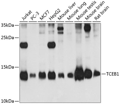
![Various whole cell extracts (30 μg) were separated by 15% SDS-PAGE, and the membrane was blotted with TCEB1 antibody [HL1683] (GTX637282) diluted at 1:50000. The HRP-conjugated anti-rabbit IgG antibody (GTX213110-01) was used to detect the primary antibody.](https://www.genetex.com/upload/website/prouct_img/normal/GTX637282/GTX637282_44881_20221230_WB_D_C_23010400_268.webp "Various whole cell extracts (30 μg) were separated by 15% SDS-PAGE, and the membrane was blotted with TCEB1 antibody [HL1683] (GTX637282) diluted at 1:50000. The HRP-conjugated anti-rabbit IgG antibody (GTX213110-01) was used to detect the primary antibody.")
![Non-transfected (–) and transfected (+) 293T whole cell extracts (30 μg) were separated by 15% SDS-PAGE, and the membrane was blotted with TCEB1 antibody [HL1683] (GTX637282) diluted at 1:500. The HRP-conjugated anti-rabbit IgG antibody (GTX213110-01) was used to detect the primary antibody.](https://www.genetex.com/upload/website/prouct_img/normal/GTX637282/GTX637282_44881_20230210_WB_shRNA_watermark_23021401_824.webp "Non-transfected (–) and transfected (+) 293T whole cell extracts (30 μg) were separated by 15% SDS-PAGE, and the membrane was blotted with TCEB1 antibody [HL1683] (GTX637282) diluted at 1:500. The HRP-conjugated anti-rabbit IgG antibody (GTX213110-01) was used to detect the primary antibody.")
![Whole cell extract (30 μg) was separated by 15% SDS-PAGE, and the membrane was blotted with TCEB1 antibody [HL1683] (GTX637282) diluted at 1:1000. The HRP-conjugated anti-rabbit IgG antibody (GTX213110-01) was used to detect the primary antibody.](https://www.genetex.com/upload/website/prouct_img/normal/GTX637282/GTX637282_44881_20230707_WB_Drosophila_23071223_722.webp "Whole cell extract (30 μg) was separated by 15% SDS-PAGE, and the membrane was blotted with TCEB1 antibody [HL1683] (GTX637282) diluted at 1:1000. The HRP-conjugated anti-rabbit IgG antibody (GTX213110-01) was used to detect the primary antibody.")
![Various whole cell extracts (30 μg) were separated by 15% SDS-PAGE, and the membrane was blotted with TCEB1 antibody [HL1683] (GTX637282) diluted at 1:50000. The HRP-conjugated anti-rabbit IgG antibody (GTX213110-01) was used to detect the primary antibody.](https://www.genetex.com/upload/website/prouct_img/normal/GTX637282/GTX637282_45125_20230804_WB_23081619_390.webp "Various whole cell extracts (30 μg) were separated by 15% SDS-PAGE, and the membrane was blotted with TCEB1 antibody [HL1683] (GTX637282) diluted at 1:50000. The HRP-conjugated anti-rabbit IgG antibody (GTX213110-01) was used to detect the primary antibody.")
![Whole cell extract (30 μg) was separated by 15% SDS-PAGE, and the membrane was blotted with TCEB1 antibody [HL1683] (GTX637282) diluted at 1:1000. The HRP-conjugated anti-rabbit IgG antibody (GTX213110-01) was used to detect the primary antibody.](https://www.genetex.com/upload/website/prouct_img/normal/GTX637282/GTX637282_45125_20250815_WB_R_25082121_638.webp "Whole cell extract (30 μg) was separated by 15% SDS-PAGE, and the membrane was blotted with TCEB1 antibody [HL1683] (GTX637282) diluted at 1:1000. The HRP-conjugated anti-rabbit IgG antibody (GTX213110-01) was used to detect the primary antibody.")
![Whole cell extract (30 μg) was separated by 15% SDS-PAGE, and the membrane was blotted with TCEB1 antibody [HL1683] (GTX637282) diluted at 1:1000. The HRP-conjugated anti-rabbit IgG antibody (GTX213110-01) was used to detect the primary antibody.](https://www.genetex.com/upload/website/prouct_img/normal/GTX637282/GTX637282_45125_20250822_WB_M_25082900_744.webp "Whole cell extract (30 μg) was separated by 15% SDS-PAGE, and the membrane was blotted with TCEB1 antibody [HL1683] (GTX637282) diluted at 1:1000. The HRP-conjugated anti-rabbit IgG antibody (GTX213110-01) was used to detect the primary antibody.")
TCEB1 antibody [HL1683] detects TCEB1 protein at cytoplasm by immunohistochemical analysis. Sample: Paraffin-embedded mouse muscle. TCEB1 stained by TCEB1 antibody [HL1683] (GTX637282) diluted at 1:100. Antigen Retrieval: Citrate buffer, pH 6.0, 15 min
TCEB1 antibody [HL1683]
GTX637282
ApplicationsWestern Blot, ImmunoHistoChemistry, ImmunoHistoChemistry Paraffin
Product group Antibodies
ReactivityCanine, Drosophila, Feline, Human, Mouse
TargetELOC
Overview
- SupplierGeneTex
- Product NameTCEB1 antibody [HL1683]
- Delivery Days Customer9
- Application Supplier NoteWB: 1:5000-1:50000. *Optimal dilutions/concentrations should be determined by the researcher.Not tested in other applications.
- ApplicationsWestern Blot, ImmunoHistoChemistry, ImmunoHistoChemistry Paraffin
- CertificationResearch Use Only
- ClonalityMonoclonal
- Clone IDHL1683
- Concentration1 mg/ml
- ConjugateUnconjugated
- Gene ID6921
- Target nameELOC
- Target descriptionelongin C
- Target synonymsSIII, TCEB1, elongin-C, RNA polymerase II transcription factor SIII subunit C, SIII p15, elongin 15 kDa subunit, transcription elongation factor B (SIII), polypeptide 1 (15kDa, elongin C), transcription elongation factor B polypeptide 1, transcription elongation factor B subunit 1
- HostRabbit
- IsotypeIgG
- Protein IDQ15369
- Protein NameElongin-C
- Scientific DescriptionThis gene encodes the protein elongin C, which is a subunit of the transcription factor B (SIII) complex. The SIII complex is composed of elongins A/A2, B and C. It activates elongation by RNA polymerase II by suppressing transient pausing of the polymerase at many sites within transcription units. Elongin A functions as the transcriptionally active component of the SIII complex, whereas elongins B and C are regulatory subunits. Elongin A2 is specifically expressed in the testis, and capable of forming a stable complex with elongins B and C. The von Hippel-Lindau tumor suppressor protein binds to elongins B and C, and thereby inhibits transcription elongation. Multiple alternatively spliced transcript variants encoding two distinct isoforms have been identified. [provided by RefSeq, Mar 2011]
- ReactivityCanine, Drosophila, Feline, Human, Mouse
- Storage Instruction-20°C or -80°C,2°C to 8°C
- UNSPSC41116161
Datasheet
Related products
Product group Antibodies
ELOC AntibodyCSB-PA023281EA01HU
ApplicationsELISA
ReactivityHuman
TargetELOC
- SizePrice

Product group Antibodies
Anti-Elongin-C/ELOC Antibody Picoband(r)A31720-1-CARRIER-FREE
ApplicationsFlow Cytometry, ImmunoFluorescence, Western Blot, ELISA, ImmunoCytoChemistry, ImmunoHistoChemistry
ReactivityHuman, Mouse, Rat
TargetELOC
- SizePrice
Product group Antibodies
Anti-TCEB1 AntibodyA31227
ApplicationsWestern Blot, ImmunoHistoChemistry
ReactivityHuman, Mouse, Rat
- SizePrice
Product group Antibodies

TCEB1 / Elongin C AntibodyLS-C766339
ApplicationsELISA, ImmunoHistoChemistry
ReactivityHuman, Mouse, Rat
TargetELOC
- SizePrice

Product group Antibodies
Anti-ELOC AntibodyHPA078113
ApplicationsImmunoCytoChemistry
ReactivityHuman
TargetELOC
- SizePrice
Product group Antibodies
ApplicationsImmunoPrecipitation, Western Blot, ImmunoCytoChemistry, ImmunoHistoChemistry
ReactivityMouse, Rat
TargetELOC
- SizePrice
Product group Antibodies
TCEB1 antibodyGTX65573
ApplicationsWestern Blot
ReactivityHuman, Mouse, Rat
TargetELOC
- SizePrice
Product group Antibodies
TCEB1 antibodyGTX132302
ApplicationsWestern Blot
ReactivityCanine, Feline, Human
TargetELOC
- SizePrice
Product group Antibodies
Anti-TCEB1 Antibody144-12515
ApplicationsWestern Blot
ReactivityHuman, Mouse, Rat
TargetELOC
- SizePrice