

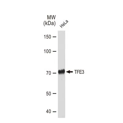
WB analysis of HeLa whole cell lysate using GTX03229 TFE3 antibody [GT1317]. Dilution : 1:1000 Loading : 25μg per lane
![IHC-P analysis of human esophageal cancer section using GTX03229 TFE3 antibody [GT1317]. Dilution : 1:100](https://www.genetex.com/upload/website/prouct_img/normal/GTX03229/GTX03229_20210615_IHC-P_1_w_23053123_778.webp "IHC-P analysis of human esophageal cancer section using GTX03229 TFE3 antibody [GT1317]. Dilution : 1:100")
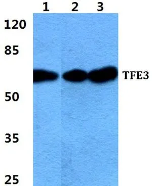
![WB analysis of various samples using GTX03229 TFE3 antibody [GT1317]. Dilution : 1:1000 Loading : 25μg per lane](https://www.genetex.com/upload/website/prouct_img/normal/GTX03229/GTX03229_1_WB_w_23053123_142.webp "WB analysis of various samples using GTX03229 TFE3 antibody [GT1317]. Dilution : 1:1000 Loading : 25μg per lane")
![Various whole cell extracts (30 μg) were separated by 7.5% SDS-PAGE, and the membrane was blotted with TFE3 antibody [GT1317] (GTX03229) diluted at 1:1000. The HRP-conjugated anti-rabbit IgG antibody (GTX213110-01) was used to detect the primary antibody.](https://www.genetex.com/upload/website/prouct_img/normal/GTX03229/GTX03229_4000001829_20210709_WB_TPM_watermark_w_23053123_824.webp "Various whole cell extracts (30 μg) were separated by 7.5% SDS-PAGE, and the membrane was blotted with TFE3 antibody [GT1317] (GTX03229) diluted at 1:1000. The HRP-conjugated anti-rabbit IgG antibody (GTX213110-01) was used to detect the primary antibody.")
WB analysis of HeLa whole cell lysate using GTX03229 TFE3 antibody [GT1317]. Dilution : 1:1000 Loading : 25μg per lane
TFE3 antibody [GT1317]
GTX03229
ApplicationsWestern Blot, ImmunoHistoChemistry, ImmunoHistoChemistry Paraffin
Product group Antibodies
ReactivityHuman, Mouse, Rat
TargetTFE3
Overview
- SupplierGeneTex
- Product NameTFE3 antibody [GT1317]
- Delivery Days Customer9
- Application Supplier NoteWB: 1:500 - 1:2000. IHC-P: 1:50 - 1:200. *Optimal dilutions/concentrations should be determined by the researcher.Not tested in other applications.
- ApplicationsWestern Blot, ImmunoHistoChemistry, ImmunoHistoChemistry Paraffin
- CertificationResearch Use Only
- ClonalityMonoclonal
- Clone IDGT1317
- Concentration0.75 mg/ml
- ConjugateUnconjugated
- Gene ID7030
- Target nameTFE3
- Target descriptiontranscription factor binding to IGHM enhancer 3
- Target synonymsMRXSPF, RCCP2, RCCX1, TFEA, bHLHe33, transcription factor E3, class E basic helix-loop-helix protein 33, transcription factor E family, member A, transcription factor for IgH enhancer, transcription factor for immunoglobulin heavy-chain enhancer 3
- HostRabbit
- IsotypeIgG
- Protein IDP19532
- Protein NameTranscription factor E3
- Scientific DescriptionThis gene encodes a basic helix-loop-helix domain-containing transcription factor that binds MUE3-type E-box sequences in the promoter of genes. The encoded protein promotes the expression of genes downstream of transforming growth factor beta (TGF-beta) signaling. This gene may be involved in chromosomal translocations in renal cell carcinomas and other cancers, resulting in the production of fusion proteins. Translocation partners include PRCC (papillary renal cell carcinoma), NONO (non-POU domain containing, octamer-binding), and ASPSCR1 (alveolar soft part sarcoma chromosome region, candidate 1), among other genes. Alternative splicing results in multiple transcript variants. [provided by RefSeq, Aug 2013]
- ReactivityHuman, Mouse, Rat
- Storage Instruction-20°C or -80°C,2°C to 8°C
- UNSPSC12352203
Datasheet
Related products
Product group Antibodies
Anti-TFE3 Antibody118-10036
ApplicationsWestern Blot, ELISA, ImmunoHistoChemistry
ReactivityHuman
- SizePrice

Product group Antibodies
Anti-TFE3 AntibodyAMAB91798
ApplicationsWestern Blot, ImmunoHistoChemistry
ReactivityHuman
TargetTFE3
- SizePrice

Product group Antibodies
ApplicationsFlow Cytometry, ImmunoFluorescence, ImmunoCytoChemistry, ImmunoHistoChemistry
ReactivityHuman
TargetTFE3
- SizePrice
![IHC-P analysis of human renal cell carcinoma (RCC) tissue using GTX04963 TFE3 antibody [MSVA-403R] HistoMAX?. Strong nuclear TFE3 positivity in a renal cell carcinoma with TFE3 fusion.](https://www.genetex.com/upload/website/prouct_img/normal/GTX04963/GTX04963_20241028_IHC-P_24102820_366.webp)
Product group Antibodies
ApplicationsImmunoHistoChemistry, ImmunoHistoChemistry Paraffin
ReactivityHuman
TargetTFE3
- SizePrice
Product group Antibodies
TFE3 antibodyGTX66746
ApplicationsWestern Blot, ImmunoHistoChemistry, ImmunoHistoChemistry Paraffin
ReactivityHuman, Mouse, Rat
TargetTFE3
- SizePrice
Product group Antibodies
TFE3 Polyclonal AntibodyCAC13217
ApplicationsWestern Blot, ELISA, ImmunoHistoChemistry
TargetTFE3
- SizePrice
Product group Antibodies
TFE3 Recombinant Antibody, Biotin ConjugatedBSM-61810R-BIOTIN
ApplicationsImmunoHistoChemistry, ImmunoHistoChemistry Frozen, ImmunoHistoChemistry Paraffin
ReactivityHuman
TargetTFE3
- SizePrice
Product group Antibodies
Anti-TFE3 AntibodyA83833
ApplicationsWestern Blot, ELISA
ReactivityHuman
- SizePrice