

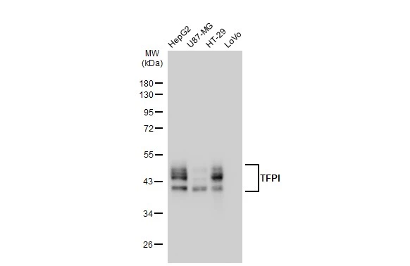
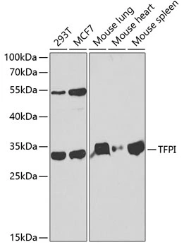
Various whole cell extracts (30 μg) were separated by 10% SDS-PAGE, and the membrane was blotted with TFPI antibody [GT1246] (GTX02843) diluted at 1:1000. The HRP-conjugated anti-rabbit IgG antibody (GTX213110-01) was used to detect the primary antibody.
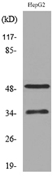
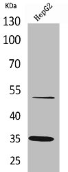
![WB analysis of BxPC-3 whole cell lysate using GTX02843 TFPI antibody [GT1246]. Dilution : 1:1000 Loading : 25μg](https://www.genetex.com/upload/website/prouct_img/normal/GTX02843/CutImage_A8704_WB_01_(1070825)_w_23053123_134.webp "WB analysis of BxPC-3 whole cell lysate using GTX02843 TFPI antibody [GT1246]. Dilution : 1:1000 Loading : 25μg")
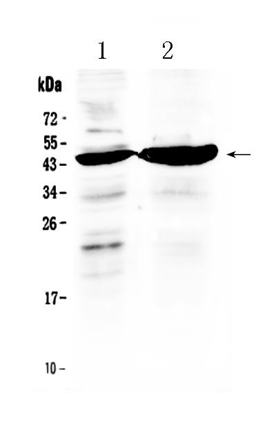
Various whole cell extracts (30 μg) were separated by 10% SDS-PAGE, and the membrane was blotted with TFPI antibody [GT1246] (GTX02843) diluted at 1:1000. The HRP-conjugated anti-rabbit IgG antibody (GTX213110-01) was used to detect the primary antibody.
TFPI antibody [GT1246]
GTX02843
ApplicationsWestern Blot
Product group Antibodies
ReactivityHuman
TargetTFPI
Overview
- SupplierGeneTex
- Product NameTFPI antibody [GT1246]
- Delivery Days Customer9
- Application Supplier NoteWB: 1:500 - 1:2000. *Optimal dilutions/concentrations should be determined by the researcher.Not tested in other applications.
- ApplicationsWestern Blot
- CertificationResearch Use Only
- ClonalityMonoclonal
- Clone IDGT1246
- Concentration0.71 mg/ml
- ConjugateUnconjugated
- Gene ID7035
- Target nameTFPI
- Target descriptiontissue factor pathway inhibitor
- Target synonymsEPI, LACI, TFI, TFPI1, tissue factor pathway inhibitor, anti-convertin, extrinsic pathway inhibitor, tissue factor pathway inhibitor (lipoprotein-associated coagulation inhibitor)
- HostRabbit
- IsotypeIgG
- Protein IDP10646
- Protein NameTissue factor pathway inhibitor
- Scientific DescriptionThis gene encodes a Kunitz-type serine protease inhibitor that regulates the tissue factor (TF)-dependent pathway of blood coagulation. The coagulation process initiates with the formation of a factor VIIa-TF complex, which proteolytically activates additional proteases (factors IX and X) and ultimately leads to the formation of a fibrin clot. The product of this gene inhibits the activated factor X and VIIa-TF proteases in an autoregulatory loop. Inhibition of the encoded protein restores hemostasis in animal models of hemophilia. This gene encodes multiple protein isoforms that differ in their inhibitory activity, specificity and cellular localization. [provided by RefSeq, Jul 2016]
- ReactivityHuman
- Storage Instruction-20°C,2°C to 8°C
- UNSPSC41116161
Datasheet
Related products
Product group Antibodies
Anti-TFPI AntibodyA101293
ApplicationsWestern Blot, ELISA
ReactivityHuman
- SizePrice
Product group Antibodies
Anti-TFPI Antibody Picoband(r)A01052-CARRIER-FREE
ApplicationsWestern Blot
ReactivityHuman
TargetTFPI
- SizePrice
Product group Antibodies
Anti-TFPI Antibody144-01624
ApplicationsImmunoFluorescence, Western Blot
ReactivityHuman, Mouse
TargetTFPI
- SizePrice
Product group Antibodies

ApplicationsELISA
ReactivityHuman
TargetTFPI
- SizePrice
Product group Antibodies
TFPI Polyclonal AntibodyBS-2535R
ApplicationsImmunoFluorescence, Western Blot, ELISA, ImmunoCytoChemistry, ImmunoHistoChemistry, ImmunoHistoChemistry Frozen, ImmunoHistoChemistry Paraffin
ReactivityBovine, Equine, Human, Mouse, Porcine, Rabbit, Rat
TargetTFPI
- SizePrice
Product group Antibodies

ApplicationsWestern Blot, ELISA
ReactivityHuman
TargetTFPI
- SizePrice
Product group Antibodies
ApplicationsImmunoPrecipitation, Western Blot, ImmunoCytoChemistry, ImmunoHistoChemistry
TargetTFPI
- SizePrice
Product group Antibodies
TFPI AntibodyCSB-PA005186
ApplicationsWestern Blot, ELISA
ReactivityHuman
TargetTFPI
- SizePrice
Product group Antibodies
TFPI antibodyGTX16395
ApplicationsImmunoFluorescence, Western Blot, ImmunoCytoChemistry
ReactivityHuman, Mouse
TargetTFPI
- SizePrice