

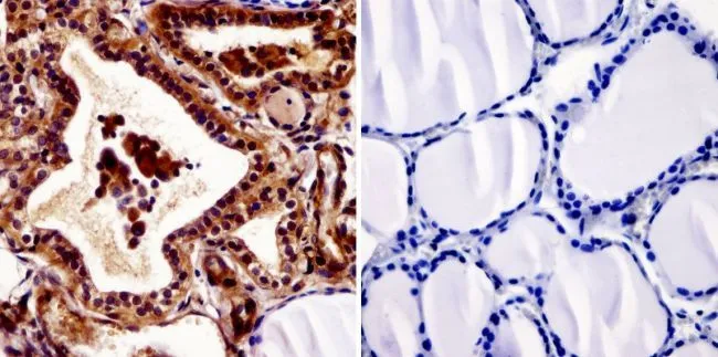
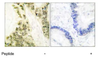
IHC-P analysis of human thyroid tissue using GTX22743 Thyroid Hormone Receptor antibody [C3]. Left : Primary antibody Right : Negative control without primary antibody Antigen retrieval : heat induced antigen retrieval was performed using 10mM sodium citrate (pH6.0) buffer, microwaved for 8-15 minutes Dilution : 1:20
![IHC-P analysis of human thyroid tissue using GTX22743 Thyroid Hormone Receptor antibody [C3]. Right : Primary antibody Left : Negative control without primary antibody Antigen retrieval : 10mM sodium citrate (pH 6.0), microwaved for 8-15 min Dilution : 1:20](https://www.genetex.com/upload/website/prouct_img/normal/GTX22743/GTX22743_1092_IHC-P_w_23060620_731.webp "IHC-P analysis of human thyroid tissue using GTX22743 Thyroid Hormone Receptor antibody [C3]. Right : Primary antibody Left : Negative control without primary antibody Antigen retrieval : 10mM sodium citrate (pH 6.0), microwaved for 8-15 min Dilution : 1:20")
![IHC-P analysis of mouse thyroid tissue using GTX22743 Thyroid Hormone Receptor antibody [C3]. Right : Primary antibody Left : Negative control without primary antibody Antigen retrieval : 10mM sodium citrate (pH 6.0), microwaved for 8-15 min Dilution : 1:20](https://www.genetex.com/upload/website/prouct_img/normal/GTX22743/GTX22743_1093_IHC-P_w_23060620_859.webp "IHC-P analysis of mouse thyroid tissue using GTX22743 Thyroid Hormone Receptor antibody [C3]. Right : Primary antibody Left : Negative control without primary antibody Antigen retrieval : 10mM sodium citrate (pH 6.0), microwaved for 8-15 min Dilution : 1:20")
![IHC-P analysis of human colon tissue using GTX22743 Thyroid Hormone Receptor antibody [C3]. Right : Primary antibody Left : Negative control without primary antibody Antigen retrieval : 10mM sodium citrate (pH 6.0), microwaved for 8-15 min Dilution : 1:20](https://www.genetex.com/upload/website/prouct_img/normal/GTX22743/GTX22743_1094_IHC-P_w_23060620_881.webp "IHC-P analysis of human colon tissue using GTX22743 Thyroid Hormone Receptor antibody [C3]. Right : Primary antibody Left : Negative control without primary antibody Antigen retrieval : 10mM sodium citrate (pH 6.0), microwaved for 8-15 min Dilution : 1:20")
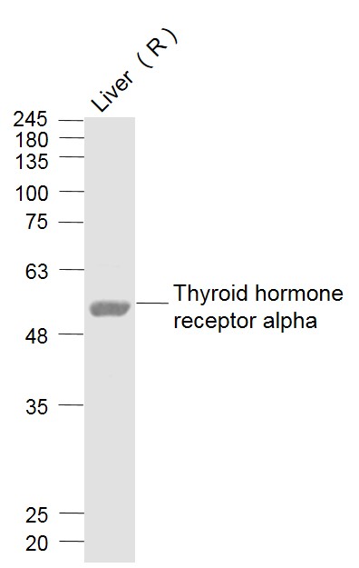
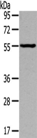
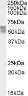
![WB analysis of samples using GTX22743 Thyroid Hormone Receptor antibody [C3].](https://www.genetex.com/upload/website/prouct_img/normal/GTX22743/GTX22743_1580_WB_w_23060620_351.webp "WB analysis of samples using GTX22743 Thyroid Hormone Receptor antibody [C3].")
![ICC/IF analysis of HeLa cells using GTX22743 Thyroid Hormone Receptor antibody [C3]. Cells were probed without (right) or with(left) an antibody. Green : Primary antibody Blue : Nuclei Red : Actin Fixation : formaldehyde Dilution : 1:20 overnight at 4oC](https://www.genetex.com/upload/website/prouct_img/normal/GTX22743/GTX22743_405_ICC-IF_w_23060620_961.webp "ICC/IF analysis of HeLa cells using GTX22743 Thyroid Hormone Receptor antibody [C3]. Cells were probed without (right) or with(left) an antibody. Green : Primary antibody Blue : Nuclei Red : Actin Fixation : formaldehyde Dilution : 1:20 overnight at 4oC")
![ICC/IF analysis of HepG2 cells using GTX22743 Thyroid Hormone Receptor antibody [C3]. Cells were probed without (right) or with(left) an antibody. Green : Primary antibody Blue : Nuclei Red : Actin Fixation : formaldehyde Dilution : 1:20 overnight at 4oC](https://www.genetex.com/upload/website/prouct_img/normal/GTX22743/GTX22743_406_ICC-IF_w_23060620_795.webp "ICC/IF analysis of HepG2 cells using GTX22743 Thyroid Hormone Receptor antibody [C3]. Cells were probed without (right) or with(left) an antibody. Green : Primary antibody Blue : Nuclei Red : Actin Fixation : formaldehyde Dilution : 1:20 overnight at 4oC")
IHC-P analysis of human thyroid tissue using GTX22743 Thyroid Hormone Receptor antibody [C3]. Left : Primary antibody Right : Negative control without primary antibody Antigen retrieval : heat induced antigen retrieval was performed using 10mM sodium citrate (pH6.0) buffer, microwaved for 8-15 minutes Dilution : 1:20
Thyroid Hormone Receptor antibody [C3]
GTX22743
ApplicationsGel Shift Assay, ImmunoFluorescence, ImmunoPrecipitation, Western Blot, ImmunoCytoChemistry, ImmunoHistoChemistry, ImmunoHistoChemistry Paraffin
Product group Antibodies
ReactivityHuman, Mouse, Primate, Rat, Xenopus
TargetTHRA
Overview
- SupplierGeneTex
- Product NameThyroid Hormone Receptor antibody [C3]
- Delivery Days Customer9
- Application Supplier NoteWB: 1:1,000. ICC/IF: 1:20. IHC-P: 1:20. *Optimal dilutions/concentrations should be determined by the researcher.Not tested in other applications.
- ApplicationsGel Shift Assay, ImmunoFluorescence, ImmunoPrecipitation, Western Blot, ImmunoCytoChemistry, ImmunoHistoChemistry, ImmunoHistoChemistry Paraffin
- CertificationResearch Use Only
- ClonalityMonoclonal
- Clone IDC3
- ConjugateUnconjugated
- Gene ID7067
- Target nameTHRA
- Target descriptionthyroid hormone receptor alpha
- Target synonymsAR7, CHNG6, EAR7, ERB-T-1, ERBA, ERBA1, NR1A1, THRA1, THRA2, THRalpha, THRalpha1, THRalpha2, TRalpha, TRalpha1, TRalpha2, c-ERBA-1, c-erbA, thyroid hormone receptor alpha, EAR-7, ERBA-related 7, V-erbA-related protein 7, c-erbA protooncogene, c-erbA-alpha, nuclear receptor subfamily 1 group A member 1, thyroid hormone receptor alpha 1, thyroid hormone receptor, alpha (erythroblastic leukemia viral (v-erb-a) oncogene homolog, avian), triiodothyronine receptor
- HostMouse
- IsotypeIgG1
- Protein IDP10827
- Protein NameThyroid hormone receptor alpha
- Scientific DescriptionThe protein encoded by this gene is a nuclear hormone receptor for triiodothyronine. It is one of the several receptors for thyroid hormone, and has been shown to mediate the biological activities of thyroid hormone. Knockout studies in mice suggest that the different receptors, while having certain extent of redundancy, may mediate different functions of thyroid hormone. Alternatively spliced transcript variants encoding distinct isoforms have been reported. [provided by RefSeq, Jul 2008]
- ReactivityHuman, Mouse, Primate, Rat, Xenopus
- Storage Instruction-20°C or -80°C,2°C to 8°C
- UNSPSC12352203
References
- Hasebe T, Fujimoto K, Buchholz DR, et al. Stem cell development involves divergent thyroid hormone receptor subtype expression and epigenetic modifications in the Xenopus metamorphosing intestine. Gen Comp Endocrinol. 2020,292:113441. doi: 10.1016/j.ygcen.2020.113441Read this paper
Datasheet
Related products
Product group Antibodies
ApplicationsFlow Cytometry, ImmunoFluorescence, Western Blot, ELISA, ImmunoCytoChemistry, ImmunoHistoChemistry, ImmunoHistoChemistry Frozen, ImmunoHistoChemistry Paraffin
ReactivityChicken, Equine, Guinea Pig, Human, Mouse, Porcine, Rabbit, Rat, Sheep
TargetTHRA
- SizePrice
Product group Antibodies
THRA Polyclonal AntibodyCAC13220
ApplicationsWestern Blot, ELISA, ImmunoHistoChemistry
TargetTHRA
- SizePrice
Product group Antibodies
Anti-THRA (N-term) Antibody102-25025
ApplicationsWestern Blot
TargetTHRA
- SizePrice
Product group Antibodies
Anti-THRA AntibodyA44150
ApplicationsWestern Blot
ReactivityHuman, Mouse
- SizePrice
Product group Antibodies

Goat anti-THRA AntibodyEB07965
ApplicationsWestern Blot, ELISA, ImmunoHistoChemistry
ReactivityBovine, Canine, Human, Mouse, Porcine, Rat
TargetTHRA
- SizePrice
![Western blot of rat hippocampal lysate showing specific immunolabeling of the ~58k TR-alpha2 protein using THRA antibody [1330] (GTX82579)](https://www.genetex.com/upload/website/prouct_img/normal/GTX82579/Thyroid-Hormone-Receptor-alpha-antibody-1330_Western-blot_GTX82579-1_w_23061322_495.webp)
Product group Antibodies
ApplicationsWestern Blot
ReactivityHuman, Rat
TargetTHRA
- SizePrice
Product group Antibodies
ApplicationsWestern Blot, ImmunoHistoChemistry, ImmunoHistoChemistry Paraffin
ReactivityHuman
TargetTHRA
- SizePrice
Product group Antibodies
ApplicationsWestern Blot, ImmunoHistoChemistry, ImmunoHistoChemistry Paraffin
ReactivityHuman
TargetTHRA
- SizePrice