

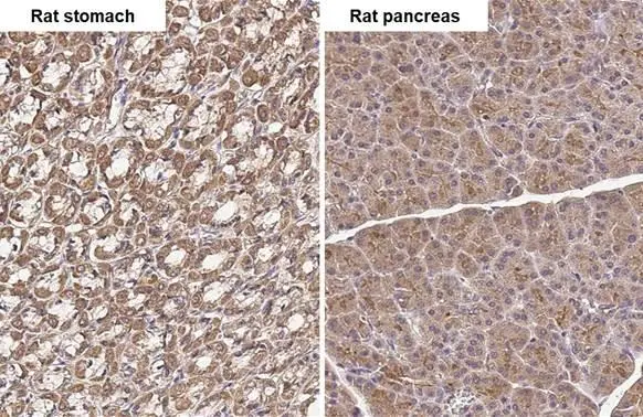
TLR3 antibody [HL2314] detects TLR3 protein by immunohistochemical analysis. Sample: Paraffin-embedded rat tissues. TLR3 stained by TLR3 antibody [HL2314] (GTX638477) diluted at 1:100. Antigen Retrieval: Citrate buffer, pH 6.0, 15 min
![TLR3 antibody [HL2314] detects TLR3 protein at cytoplasm by immunohistochemical analysis. Sample: Paraffin-embedded mouse intestine. TLR3 stained by TLR3 antibody [HL2314] (GTX638477) diluted at 1:100. Antigen Retrieval: Citrate buffer, pH 6.0, 15 min](https://www.genetex.com/upload/website/prouct_img/normal/GTX638477/GTX638477_T-45005_20230428_IHC-P_M_23050918_455.webp "TLR3 antibody [HL2314] detects TLR3 protein at cytoplasm by immunohistochemical analysis. Sample: Paraffin-embedded mouse intestine. TLR3 stained by TLR3 antibody [HL2314] (GTX638477) diluted at 1:100. Antigen Retrieval: Citrate buffer, pH 6.0, 15 min")
![Various whole cell extracts (30 μg) were separated by 7.5% SDS-PAGE, and the membrane was blotted with TLR3 antibody [HL2314] (GTX638477) diluted at 1:1000. The HRP-conjugated anti-rabbit IgG antibody (GTX213110-01) was used to detect the primary antibody.](https://www.genetex.com/upload/website/prouct_img/normal/GTX638477/GTX638477_T-45005_20230616_WB_D_C_23062019_668.webp "Various whole cell extracts (30 μg) were separated by 7.5% SDS-PAGE, and the membrane was blotted with TLR3 antibody [HL2314] (GTX638477) diluted at 1:1000. The HRP-conjugated anti-rabbit IgG antibody (GTX213110-01) was used to detect the primary antibody.")
![Various whole cell extracts (30 μg) were separated by 7.5% SDS-PAGE, and the membrane was blotted with TLR3 antibody [HL2314] (GTX638477) diluted at 1:1000. The HRP-conjugated anti-rabbit IgG antibody (GTX213110-01) was used to detect the primary antibody, and the signal was developed with Trident femto Western HRP Substrate. Corresponding RNA expression data for the same cell lines are based on Human Protein Atlas program.](https://www.genetex.com/upload/website/prouct_img/normal/GTX638477/GTX638477_45082_20230707_WB_TPM_watermark_23071223_813.webp "Various whole cell extracts (30 μg) were separated by 7.5% SDS-PAGE, and the membrane was blotted with TLR3 antibody [HL2314] (GTX638477) diluted at 1:1000. The HRP-conjugated anti-rabbit IgG antibody (GTX213110-01) was used to detect the primary antibody, and the signal was developed with Trident femto Western HRP Substrate. Corresponding RNA expression data for the same cell lines are based on Human Protein Atlas program.")
![TLR3 antibody [HL2314] detects TLR3 protein at cytoplasm by immunohistochemical analysis. Sample: Paraffin-embedded dog brain. TLR3 stained by TLR3 antibody [HL2314] (GTX638477) diluted at 1:100. Antigen Retrieval: Citrate buffer, pH 6.0, 15 min](https://www.genetex.com/upload/website/prouct_img/normal/GTX638477/GTX638477_45082_20230804_IHC-P_Dog_23090619_949.webp "TLR3 antibody [HL2314] detects TLR3 protein at cytoplasm by immunohistochemical analysis. Sample: Paraffin-embedded dog brain. TLR3 stained by TLR3 antibody [HL2314] (GTX638477) diluted at 1:100. Antigen Retrieval: Citrate buffer, pH 6.0, 15 min")
TLR3 antibody [HL2314] detects TLR3 protein by immunohistochemical analysis. Sample: Paraffin-embedded rat tissues. TLR3 stained by TLR3 antibody [HL2314] (GTX638477) diluted at 1:100. Antigen Retrieval: Citrate buffer, pH 6.0, 15 min
TLR3 antibody [HL2314]
GTX638477
ApplicationsWestern Blot, ImmunoHistoChemistry, ImmunoHistoChemistry Paraffin
Product group Antibodies
ReactivityCanine, Feline, Human, Mouse, Rat
TargetTLR3
Overview
- SupplierGeneTex
- Product NameTLR3 antibody [HL2314]
- Delivery Days Customer9
- Application Supplier NoteWB: 1:500-1:3000. *Optimal dilutions/concentrations should be determined by the researcher.Not tested in other applications.
- ApplicationsWestern Blot, ImmunoHistoChemistry, ImmunoHistoChemistry Paraffin
- CertificationResearch Use Only
- ClonalityMonoclonal
- Clone IDHL2314
- Concentration1 mg/ml
- ConjugateUnconjugated
- Gene ID7098
- Target nameTLR3
- Target descriptiontoll like receptor 3
- Target synonymsCD283, IIAE2, IMD83, toll-like receptor 3
- HostRabbit
- IsotypeIgG
- Protein IDO15455
- Protein NameToll-like receptor 3
- Scientific DescriptionThe protein encoded by this gene is a member of the Toll-like receptor (TLR) family which plays a fundamental role in pathogen recognition and activation of innate immunity. TLRs are highly conserved from Drosophila to humans and share structural and functional similarities. They recognize pathogen-associated molecular patterns (PAMPs) that are expressed on infectious agents, and mediate the production of cytokines necessary for the development of effective immunity. The various TLRs exhibit different patterns of expression. This receptor is most abundantly expressed in placenta and pancreas, and is restricted to the dendritic subpopulation of the leukocytes. It recognizes dsRNA associated with viral infection, and induces the activation of NF-kappaB and the production of type I interferons. It may thus play a role in host defense against viruses. Use of alternative polyadenylation sites to generate different length transcripts has been noted for this gene. [provided by RefSeq, Jul 2008]
- ReactivityCanine, Feline, Human, Mouse, Rat
- Storage Instruction-20°C or -80°C,2°C to 8°C
- UNSPSC41116161
Datasheet
Related products
Product group Antibodies
Anti-TLR3 Antibody144-62136
ApplicationsImmunoFluorescence, Western Blot
ReactivityHuman, Mouse, Rat
TargetTLR3
- SizePrice

Product group Antibodies
Anti-TLR3 Antibody Picoband(r)A00197-CARRIER-FREE
ApplicationsWestern Blot, ELISA
ReactivityHuman, Mouse, Rat
TargetTLR3
- SizePrice
Product group Antibodies
TLR3 AntibodyCSB-PA023602LA01HU
ApplicationsImmunoFluorescence, ELISA, ImmunoHistoChemistry
ReactivityHuman
TargetTLR3
- SizePrice
Product group Antibodies
ApplicationsImmunoPrecipitation, Western Blot, ImmunoCytoChemistry, ImmunoHistoChemistry
TargetTLR3
- SizePrice
![IHC-P analysis of mouse colon tissue using GTX13915 TLR3 antibody [40C1285.6]. Dilution : 1:500](https://www.genetex.com/upload/website/prouct_img/normal/GTX13915/GTX13915_442_IHC-P_w_23060620_693.webp)
Product group Antibodies
TLR3 antibody [40C1285.6]GTX13915
ApplicationsFlow Cytometry, ImmunoFluorescence, ImmunoPrecipitation, Western Blot, ImmunoCytoChemistry, ImmunoHistoChemistry, ImmunoHistoChemistry Paraffin
ReactivityCanine, Human, Mouse
TargetTLR3
- SizePrice
Product group Antibodies
References
TLR3 antibodyGTX20260
ApplicationsImmunoFluorescence, Western Blot, ELISA, ImmunoCytoChemistry, ImmunoHistoChemistry, ImmunoHistoChemistry Paraffin
ReactivityHuman, Mouse
TargetTLR3
- SizePrice
Product group Antibodies
TLR3 antibodyGTX31291
ApplicationsImmunoFluorescence, Western Blot, ELISA, ImmunoCytoChemistry
ReactivityHuman, Mouse
TargetTLR3
- SizePrice
Product group Antibodies
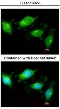
TLR3 antibodyGTX113022
ApplicationsImmunoFluorescence, Western Blot, ImmunoCytoChemistry, ImmunoHistoChemistry, ImmunoHistoChemistry Paraffin
ReactivityHuman, Mouse, Rat
TargetTLR3
- SizePrice
Product group Antibodies

TLR3 AntibodyLS-C400482
ApplicationsWestern Blot, ELISA
ReactivityHuman, Mouse
TargetTLR3
- SizePrice