

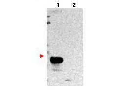
Western blot using GeneTex protein A purified anti-TMBIM1 antibody (GTX48815) shows detection of exogenous TMBIM1 in lysates from HeLa cells transfected with pcDNA3-hTMBIM1 (lane 1). No staining is observed in lysates from mock transformed HeLa cells (lane 2). To date this antibody has shown the ability to recognize overexpressed TMBIM1 but not endogenous protein. The membrane was probed with the primary antibody at a 1:1,000 dilution at 4o C, overnight.
Western blot using GeneTex protein A purified anti-TMBIM1 antibody (GTX48815) shows detection of exogenous TMBIM1 in lysates from HeLa cells transfected with pcDNA3-hTMBIM1 (lane 1). No staining is observed in lysates from mock transformed HeLa cells (lane 2). To date this antibody has shown the ability to recognize overexpressed TMBIM1 but not endogenous protein. The membrane was probed with the primary antibody at a 1:1,000 dilution at 4o C, overnight.
Tmbim1 antibody
GTX48815
ApplicationsWestern Blot, ELISA, ImmunoHistoChemistry, ImmunoHistoChemistry Paraffin
Product group Antibodies
ReactivityHuman
TargetTMBIM1
Overview
- SupplierGeneTex
- Product NameTmbim1 antibody
- Delivery Days Customer9
- Application Supplier NoteWB: 1:500-1:2000. IHC-P: 10 microg/mL. ELISA: 1:10000. *Optimal dilutions/concentrations should be determined by the researcher.Not tested in other applications.
- ApplicationsWestern Blot, ELISA, ImmunoHistoChemistry, ImmunoHistoChemistry Paraffin
- CertificationResearch Use Only
- ClonalityPolyclonal
- Concentration1.2 mg/ml
- ConjugateUnconjugated
- Gene ID64114
- Target nameTMBIM1
- Target descriptiontransmembrane BAX inhibitor motif containing 1
- Target synonymsLFG3, MST100, MSTP100, PP1201, RECS1, protein lifeguard 3, transmembrane BAX inhibitor motif-containing protein 1
- HostRabbit
- IsotypeIgG
- Protein IDQ969X1
- Protein NameProtein lifeguard 3
- Scientific DescriptionTMBIM1 (Transmicroembrane BAX inhibitor microotif-containing protein 1) is a member of the 7 TMS (7 transmembrane domains) family of receptors known to mediate the activation of various transcription factors. TMBIM1 is identified as a novel modulator of NF-kB activation. A drosophila homolog of this protein (dNMDA1 with 40% homology to hTMBIM1) is reported to be unregulated during aging and oxidative stress.
- ReactivityHuman
- Storage Instruction-20°C or -80°C,2°C to 8°C
- UNSPSC41116161
Datasheet
Related products
Product group Antibodies
TMBIM1 AntibodyCSB-PA003955
ApplicationsWestern Blot, ELISA, ImmunoHistoChemistry
ReactivityHuman, Mouse, Rat
TargetTMBIM1
- SizePrice

Product group Antibodies
Anti-TMBIM1 Antibody Picoband(r)A10501-3-CARRIER-FREE
ApplicationsFlow Cytometry, Western Blot, ImmunoHistoChemistry
ReactivityHuman, Mouse, Rat
TargetTMBIM1
- SizePrice
Product group Antibodies
Anti-TMBIM1 AntibodyA96712
ApplicationsWestern Blot, ELISA
ReactivityHuman, Mouse, Rat
- SizePrice


Product group Antibodies
Anti-TMBIM1 AntibodyHPA012093
ApplicationsWestern Blot, ImmunoHistoChemistry
ReactivityHuman, Rat
TargetTMBIM1
- SizePrice
Product group Antibodies

TMBIM1 AntibodyLS-C409785
ApplicationsWestern Blot
ReactivityHuman, Mouse, Rat
TargetTMBIM1
- SizePrice
Product group Antibodies
Tmbim1 Polyclonal AntibodyCAC09650
ApplicationsImmunoFluorescence, Western Blot, ELISA
ReactivityMouse
TargetTMBIM1
- SizePrice

Product group Antibodies
TMBIM1 antibodyGTX87296
ApplicationsWestern Blot
ReactivityHuman
TargetTMBIM1
- SizePrice
Product group Antibodies
Anti-TMBIM1 (N-term) Antibody102-21378
ApplicationsWestern Blot, ImmunoHistoChemistry, ImmunoHistoChemistry Paraffin
TargetTMBIM1
- SizePrice