

Immunohistochemical analysis of paraffin-embedded DLD-1 xenograft, using TMED9(GTX102163) antibody at 1:500 dilution.
Antigen Retrieval: Citrate buffer, pH 6.0, 15 min
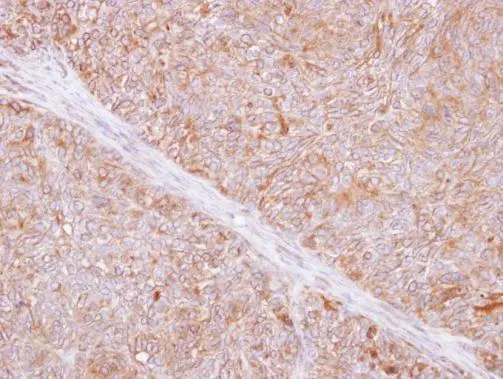
![TMED9 antibody [N1C3] detects TMED9 protein at cytoplasm in mouse duodenum by immunohistochemical analysis. Sample: Paraffin-embedded mouse duodenum. TMED9 antibody [N1C3] (GTX102163) diluted at 1:500.
Antigen Retrieval: Citrate buffer, pH 6.0, 15 min](https://www.genetex.com/upload/website/prouct_img/normal/GTX102163/GTX102163_40121_20150529_IHC_M_w_23060100_526.webp "TMED9 antibody [N1C3] detects TMED9 protein at cytoplasm in mouse duodenum by immunohistochemical analysis. Sample: Paraffin-embedded mouse duodenum. TMED9 antibody [N1C3] (GTX102163) diluted at 1:500.
Antigen Retrieval: Citrate buffer, pH 6.0, 15 min")
![MCF-7 whole cell and membrane extracts (30 μg) were separated by 12% SDS-PAGE, and the membrane was blotted with TMED9 antibody [N1C3] (GTX102163) diluted at 1:1000. The HRP-conjugated anti-rabbit IgG antibody (GTX213110-01) was used to detect the primary antibody.](https://www.genetex.com/upload/website/prouct_img/normal/GTX102163/GTX102163_45161_20230908_WB_Fraction_24041019_566.webp "MCF-7 whole cell and membrane extracts (30 μg) were separated by 12% SDS-PAGE, and the membrane was blotted with TMED9 antibody [N1C3] (GTX102163) diluted at 1:1000. The HRP-conjugated anti-rabbit IgG antibody (GTX213110-01) was used to detect the primary antibody.")
Immunohistochemical analysis of paraffin-embedded DLD-1 xenograft, using TMED9(GTX102163) antibody at 1:500 dilution.
Antigen Retrieval: Citrate buffer, pH 6.0, 15 min
TMED9 antibody [N1C3]
GTX102163
ApplicationsWestern Blot, ImmunoHistoChemistry, ImmunoHistoChemistry Paraffin
Product group Antibodies
ReactivityHuman, Mouse
TargetTMED9
Overview
- SupplierGeneTex
- Product NameTMED9 antibody [N1C3]
- Delivery Days Customer9
- Application Supplier NoteWB: 1:1000-1:10000. IHC-P: 1:100-1:1000. *Optimal dilutions/concentrations should be determined by the researcher.Not tested in other applications.
- ApplicationsWestern Blot, ImmunoHistoChemistry, ImmunoHistoChemistry Paraffin
- CertificationResearch Use Only
- ClonalityPolyclonal
- Concentration1.39 mg/ml
- ConjugateUnconjugated
- Gene ID54732
- Target nameTMED9
- Target descriptiontransmembrane p24 trafficking protein 9
- Target synonymsGMP25, HSGP25L2G, p24a2, p24alpha2, p25, transmembrane emp24 domain-containing protein 9, glycoprotein 25L2, p24 family protein alpha-2, transmembrane emp24 protein transport domain containing 9
- HostRabbit
- IsotypeIgG
- Protein IDQ9BVK6
- Protein NameTransmembrane emp24 domain-containing protein 9
- ReactivityHuman, Mouse
- Storage Instruction-20°C or -80°C,2°C to 8°C
- UNSPSC41116161
Datasheet
Related products
Product group Antibodies
Anti-TMED9 Antibody144-61366
ApplicationsWestern Blot
ReactivityHuman, Mouse, Rat
TargetTMED9
- SizePrice
Product group Antibodies
Anti-TMED9 AntibodyA88998
ApplicationsWestern Blot
ReactivityHuman, Mouse, Rat
- SizePrice
Product group Antibodies
TMED9 Polyclonal AntibodyCAC14067
ApplicationsWestern Blot, ELISA
ReactivityMouse
TargetTMED9
- SizePrice
Product group Antibodies
TMED9 AntibodyCSB-PA04804A0RB
ApplicationsWestern Blot, ELISA
ReactivityHuman, Mouse
TargetTMED9
- SizePrice
Product group Antibodies

TMED9 AntibodyLS-C332573
ApplicationsWestern Blot
ReactivityHuman, Mouse, Rat
TargetTMED9
- SizePrice
Product group Antibodies
TMED9 antibodyGTX66425
ApplicationsWestern Blot
ReactivityHuman, Mouse, Rat
TargetTMED9
- SizePrice

Product group Antibodies
Anti-TMED9 AntibodyHPA014650
ApplicationsWestern Blot, ImmunoHistoChemistry
ReactivityHuman, Rat
TargetTMED9
- SizePrice
Product group Antibodies
Anti-TMED9 AntibodyCAB3442
ApplicationsWestern Blot, ELISA
ReactivityHuman
TargetTMED9
- SizePrice