

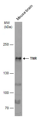
Mouse tissue extract (50 μg) was separated by 5% SDS-PAGE, and the membrane was blotted with TNR antibody [C1C2], Internal (GTX118246) diluted at 1:500.
![TNR antibody [C1C2], Internal detects TNR protein by immunofluorescent analysis. Sample: DIV9 rat E18 primary hippocampal neuron cells were fixed in 4% paraformaldehyde at RT for 15 min. Green: TNR stained by TNR antibody [C1C2], Internal (GTX118246) diluted at 1:500. Red: beta Tubulin 3/ Tuj1, stained by beta Tubulin 3/ Tuj1 antibody [GT11710] (GTX631836) diluted at 1:500. Blue: Fluoroshield with DAPI (GTX30920).](https://www.genetex.com/upload/website/prouct_img/normal/GTX118246/GTX118246_40310_20181115_ICC_IF_R_w_23060519_720.webp "TNR antibody [C1C2], Internal detects TNR protein by immunofluorescent analysis. Sample: DIV9 rat E18 primary hippocampal neuron cells were fixed in 4% paraformaldehyde at RT for 15 min. Green: TNR stained by TNR antibody [C1C2], Internal (GTX118246) diluted at 1:500. Red: beta Tubulin 3/ Tuj1, stained by beta Tubulin 3/ Tuj1 antibody [GT11710] (GTX631836) diluted at 1:500. Blue: Fluoroshield with DAPI (GTX30920).")
Mouse tissue extract (50 μg) was separated by 5% SDS-PAGE, and the membrane was blotted with TNR antibody [C1C2], Internal (GTX118246) diluted at 1:500.
TNR antibody [C1C2], Internal
GTX118246
ApplicationsImmunoFluorescence, Western Blot, ImmunoCytoChemistry
Product group Antibodies
ReactivityHuman, Mouse, Rat
TargetTNR
Overview
- SupplierGeneTex
- Product NameTNR antibody [C1C2], Internal
- Delivery Days Customer9
- Application Supplier NoteWB: 1:500-1:3000. ICC/IF: 1:100-1:1000. *Optimal dilutions/concentrations should be determined by the researcher.Not tested in other applications.
- ApplicationsImmunoFluorescence, Western Blot, ImmunoCytoChemistry
- CertificationResearch Use Only
- ClonalityPolyclonal
- Concentration0.64 mg/ml
- ConjugateUnconjugated
- Gene ID7143
- Target nameTNR
- Target descriptiontenascin R
- Target synonymsNEDSTO, TN-R, tenascin-R, janusin, restrictin
- HostRabbit
- IsotypeIgG
- Protein IDQ92752
- Protein NameTenascin-R
- Scientific DescriptionTenascin-R (TNR) is an extracellular matix protein expressed primarily in the central nervous system. It is a member of the tenascin (TN) gene family, which includes at least 3 genes in mammals: TNC (or hexabrachion; MIM 187380), TNX (TNXB; MIM 600985), and TNR (Erickson, 1993 [PubMed 7694605]). The genes are expressed in distinct tissues at different times during embryonic development and are present in adult tissues.[supplied by OMIM]
- ReactivityHuman, Mouse, Rat
- Storage Instruction-20°C or -80°C,2°C to 8°C
- UNSPSC41116161
Datasheet
Related products

Product group Antibodies
Anti-TNR AntibodyA88273
ApplicationsImmunoFluorescence, Western Blot, ImmunoCytoChemistry
ReactivityHuman, Mouse, Rat
- SizePrice

Product group Antibodies
Anti-Tenascin-R/TNR Antibody Picoband(r)A04810-1-CARRIER-FREE
ApplicationsWestern Blot, ELISA
ReactivityHuman, Mouse, Rat
TargetTNR
- SizePrice
Product group Antibodies
Anti-TNR Antibody144-60996
ApplicationsWestern Blot
ReactivityHuman, Mouse, Rat
TargetTNR
- SizePrice

Product group Antibodies
TNR AntibodyCSB-PA073278
ApplicationsELISA, ImmunoHistoChemistry
ReactivityHuman, Mouse, Rat
TargetTNR
- SizePrice
Product group Antibodies

TNR / Tenascin R AntibodyLS-C402814
ApplicationsELISA, ImmunoHistoChemistry
ReactivityHuman, Mouse, Rat
TargetTNR
- SizePrice

Product group Antibodies
Anti-TNR AntibodyHPA027134
ApplicationsImmunoHistoChemistry
ReactivityHuman
TargetTNR
- SizePrice
Product group Antibodies
TNR antibodyGTX66358
ApplicationsWestern Blot
ReactivityHuman, Mouse, Rat
TargetTNR
- SizePrice