

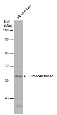
Mouse tissue extract (50 μg) was separated by 7.5% SDS-PAGE, and the membrane was blotted with Transketolase antibody [N3C2], Internal (GTX101477) diluted at 1:1000.
 A: Raji 7.5% SDS PAGE GTX101477 diluted at 1:1000")
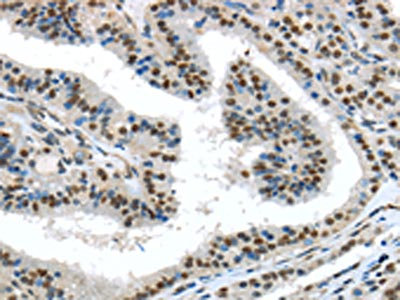
![Transketolase antibody [N3C2], Internal detects Transketolase protein at cytoplasm in mouse liver by immunohistochemical analysis. Sample: Paraffin-embedded mouse liver. Transketolase antibody [N3C2], Internal (GTX101477) diluted at 1:500.
Antigen Retrieval: Citrate buffer, pH 6.0, 15 min](https://www.genetex.com/upload/website/prouct_img/normal/GTX101477/GTX101477_40604_20160316_IHC-P_M_w_23060100_644.webp "Transketolase antibody [N3C2], Internal detects Transketolase protein at cytoplasm in mouse liver by immunohistochemical analysis. Sample: Paraffin-embedded mouse liver. Transketolase antibody [N3C2], Internal (GTX101477) diluted at 1:500.
Antigen Retrieval: Citrate buffer, pH 6.0, 15 min")
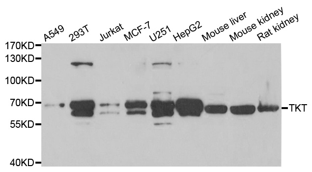
![Various whole cell extracts (30 μg) were separated by 7.5% SDS-PAGE, and the membrane was blotted with Transketolase antibody [N3C2], Internal (GTX101477) diluted at 1:1000.](https://www.genetex.com/upload/website/prouct_img/normal/GTX101477/GTX101477_40604_20160121_WB_w_23060100_562.webp "Various whole cell extracts (30 μg) were separated by 7.5% SDS-PAGE, and the membrane was blotted with Transketolase antibody [N3C2], Internal (GTX101477) diluted at 1:1000.")
 antibody at 1:500 dilution.")
Mouse tissue extract (50 μg) was separated by 7.5% SDS-PAGE, and the membrane was blotted with Transketolase antibody [N3C2], Internal (GTX101477) diluted at 1:1000.
Transketolase antibody [N3C2], Internal
GTX101477
ApplicationsImmunoFluorescence, Western Blot, ImmunoCytoChemistry, ImmunoHistoChemistry, ImmunoHistoChemistry Paraffin
Product group Antibodies
ReactivityHuman, Mouse
TargetTKT
Overview
- SupplierGeneTex
- Product NameTransketolase antibody [N3C2], Internal
- Delivery Days Customer9
- Application Supplier NoteWB: 1:500-1:3000. ICC/IF: 1:100-1:1000. IHC-P: 1:100-1:1000. *Optimal dilutions/concentrations should be determined by the researcher.Not tested in other applications.
- ApplicationsImmunoFluorescence, Western Blot, ImmunoCytoChemistry, ImmunoHistoChemistry, ImmunoHistoChemistry Paraffin
- CertificationResearch Use Only
- ClonalityPolyclonal
- Concentration1 mg/ml
- ConjugateUnconjugated
- Gene ID7086
- Target nameTKT
- Target descriptiontransketolase
- Target synonymsHEL-S-48, HEL107, SDDHD, TK, TKT1, transketolase, epididymis luminal protein 107, epididymis secretory protein Li 48
- HostRabbit
- IsotypeIgG
- Protein IDP29401
- Protein NameTransketolase
- Scientific DescriptionThis gene encodes a thiamine-dependent enzyme which plays a role in the channeling of excess sugar phosphates to glycolysis in the pentose phosphate pathway. Multiple alternatively spliced variants, encoding the same protein, have been identified. [provided by RefSeq]
- ReactivityHuman, Mouse
- Storage Instruction-20°C or -80°C,2°C to 8°C
- UNSPSC41116161
Datasheet
Related products
Product group Antibodies
Anti-TKT AntibodyA31290
ApplicationsWestern Blot, ImmunoHistoChemistry
ReactivityHuman, Mouse, Rat
- SizePrice
Product group Antibodies
Anti-TKT Antibody144-06314
ApplicationsImmunoFluorescence, Western Blot
ReactivityHuman, Mouse, Rat
TargetTKT
- SizePrice
Product group Antibodies

TKT / Transketolase AntibodyLS-C748598
ApplicationsWestern Blot, ImmunoHistoChemistry
ReactivityHuman, Mouse, Rat
TargetTKT
- SizePrice

Product group Antibodies
Anti-Transketolase/TKT Antibody Picoband(r)A02197-1-CARRIER-FREE
ApplicationsFlow Cytometry, ImmunoFluorescence, Western Blot, ELISA, ImmunoCytoChemistry, ImmunoHistoChemistry
ReactivityHuman, Mouse, Rat
TargetTKT
- SizePrice
Product group Antibodies
ApplicationsImmunoFluorescence, ELISA, ImmunoHistoChemistry, ImmunoHistoChemistry Frozen, ImmunoHistoChemistry Paraffin
ReactivityPlant
TargetTKT
- SizePrice
Product group Antibodies
ApplicationsWestern Blot, ImmunoHistoChemistry
TargetTKT
- SizePrice
Product group Antibodies
TKT AntibodyCSB-PA025245
ApplicationsELISA, ImmunoHistoChemistry
ReactivityHuman, Mouse, Rat
TargetTKT
- SizePrice
Product group Antibodies
Transketolase antibodyGTX16442
ApplicationsWestern Blot, ImmunoHistoChemistry, ImmunoHistoChemistry Paraffin
ReactivityHuman, Mouse, Rat
TargetTKT
- SizePrice
![FACS analysis of HeLa cells using GTX83510 Transketolase antibody [5H3]. Red : Primary antibody Blue : Negative control antibody](https://www.genetex.com/upload/website/prouct_img/normal/GTX83510/GTX83510_49_FACS_w_23061419_365.webp)
Product group Antibodies
Transketolase antibody [5H3]GTX83510
ApplicationsFlow Cytometry, ImmunoFluorescence, Western Blot, ImmunoCytoChemistry
ReactivityCanine, Human, Monkey, Mouse, Rat
TargetTKT
- SizePrice

Product group Antibodies
Anti-TKT AntibodyHPA029480
ApplicationsWestern Blot, ImmunoCytoChemistry, ImmunoHistoChemistry
ReactivityHuman, Mouse, Rat
TargetTKT
- SizePrice