

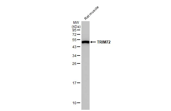
Rat tissue extract (50 μg) was separated by 12% SDS-PAGE, and the membrane was blotted with TRIM72 antibody [HL1853] (GTX637578) diluted at 1:5000. The HRP-conjugated anti-rabbit IgG antibody (GTX213110-01) was used to detect the primary antibody.
![Various tissue extracts (50 μg) were separated by 10% SDS-PAGE, and the membrane was blotted with TRIM72 antibody [HL1853] (GTX637578) diluted at 1:1000. The HRP-conjugated anti-rabbit IgG antibody (GTX213110-01) was used to detect the primary antibody.](https://www.genetex.com/upload/website/prouct_img/normal/GTX637578/GTX637578_T-44823_20221118_WB_M_tissue_22112219_448.webp "Various tissue extracts (50 μg) were separated by 10% SDS-PAGE, and the membrane was blotted with TRIM72 antibody [HL1853] (GTX637578) diluted at 1:1000. The HRP-conjugated anti-rabbit IgG antibody (GTX213110-01) was used to detect the primary antibody.")
![Various whole cell extracts (30 μg) were separated by 12% SDS-PAGE, and the membrane was blotted with TRIM72 antibody [HL1853] (GTX637578) diluted at 1:1000. The HRP-conjugated anti-rabbit IgG antibody (GTX213110-01) was used to detect the primary antibody. Corresponding RNA expression data for the same cell lines are based on Human Protein Atlas program.](https://www.genetex.com/upload/website/prouct_img/normal/GTX637578/GTX637578_44879_20221202_WB_TPM_watermark_22120719_546.webp "Various whole cell extracts (30 μg) were separated by 12% SDS-PAGE, and the membrane was blotted with TRIM72 antibody [HL1853] (GTX637578) diluted at 1:1000. The HRP-conjugated anti-rabbit IgG antibody (GTX213110-01) was used to detect the primary antibody. Corresponding RNA expression data for the same cell lines are based on Human Protein Atlas program.")
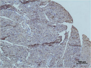
![TRIM72 antibody [HL1853] detects TRIM72 protein at cell membrane by immunohistochemical analysis. Sample: Paraffin-embedded rat tissues. TRIM72 stained by TRIM72 antibody [HL1853] (GTX637578) diluted at 1:100. Antigen Retrieval: Citrate buffer, pH 6.0, 15 min](https://www.genetex.com/upload/website/prouct_img/normal/GTX637578/GTX637578_T-44823_20221028_IHC-P_R_22122722_691.webp "TRIM72 antibody [HL1853] detects TRIM72 protein at cell membrane by immunohistochemical analysis. Sample: Paraffin-embedded rat tissues. TRIM72 stained by TRIM72 antibody [HL1853] (GTX637578) diluted at 1:100. Antigen Retrieval: Citrate buffer, pH 6.0, 15 min")
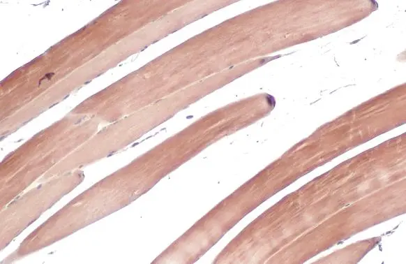
![TRIM72 antibody [HL1853] detects TRIM72 protein at cell membrane by immunohistochemical analysis. Sample: Paraffin-embedded mouse tissues. TRIM72 stained by TRIM72 antibody [HL1853] (GTX637578) diluted at 1:100. Antigen Retrieval: Citrate buffer, pH 6.0, 15 min](https://www.genetex.com/upload/website/prouct_img/normal/GTX637578/GTX637578_T-44823_20221028_IHC-P_M_22122722_923.webp "TRIM72 antibody [HL1853] detects TRIM72 protein at cell membrane by immunohistochemical analysis. Sample: Paraffin-embedded mouse tissues. TRIM72 stained by TRIM72 antibody [HL1853] (GTX637578) diluted at 1:100. Antigen Retrieval: Citrate buffer, pH 6.0, 15 min")
![Various whole cell extracts (10 μg) were separated by 12% SDS-PAGE, and the membrane was blotted with TRIM72 antibody [HL1853] (GTX637578) diluted at 1:5000. The HRP-conjugated anti-rabbit IgG antibody (GTX213110-01) was used to detect the primary antibody.](https://www.genetex.com/upload/website/prouct_img/normal/GTX637578/GTX637578_T-44823_20230113_WB_M_Differentiated_23013122_348.webp "Various whole cell extracts (10 μg) were separated by 12% SDS-PAGE, and the membrane was blotted with TRIM72 antibody [HL1853] (GTX637578) diluted at 1:5000. The HRP-conjugated anti-rabbit IgG antibody (GTX213110-01) was used to detect the primary antibody.")
Rat tissue extract (50 μg) was separated by 12% SDS-PAGE, and the membrane was blotted with TRIM72 antibody [HL1853] (GTX637578) diluted at 1:5000. The HRP-conjugated anti-rabbit IgG antibody (GTX213110-01) was used to detect the primary antibody.
TRIM72 antibody [HL1853]
GTX637578
ApplicationsWestern Blot, ImmunoHistoChemistry, ImmunoHistoChemistry Paraffin
Product group Antibodies
ReactivityHuman, Mouse, Rat
TargetTRIM72
Overview
- SupplierGeneTex
- Product NameTRIM72 antibody [HL1853]
- Delivery Days Customer9
- Application Supplier NoteWB: 1:500-1:3000. *Optimal dilutions/concentrations should be determined by the researcher.Not tested in other applications.
- ApplicationsWestern Blot, ImmunoHistoChemistry, ImmunoHistoChemistry Paraffin
- CertificationResearch Use Only
- ClonalityMonoclonal
- Clone IDHL1853
- Concentration1 mg/ml
- ConjugateUnconjugated
- Gene ID493829
- Target nameTRIM72
- Target descriptiontripartite motif containing 72
- Target synonymsMG53, tripartite motif-containing protein 72, mitsugumin-53, tripartite motif containing 72, E3 ubiquitin protein ligase
- HostRabbit
- IsotypeIgG
- Protein IDQ6ZMU5
- Protein NameTripartite motif-containing protein 72
- Scientific DescriptionEnables identical protein binding activity. Predicted to be involved in several processes, including cellular protein metabolic process; plasma membrane repair; and protein homooligomerization. Predicted to act upstream of or within negative regulation of insulin receptor signaling pathway; negative regulation of insulin-like growth factor receptor signaling pathway; and negative regulation of myotube differentiation. Predicted to be located in cytoplasmic vesicle membrane. Predicted to be active in cytoplasm and sarcolemma. [provided by Alliance of Genome Resources, Apr 2022]
- ReactivityHuman, Mouse, Rat
- Storage Instruction-20°C or -80°C,2°C to 8°C
- UNSPSC41116161
Datasheet
Related products
Product group Antibodies

Anti-MG53 [#84]Ab02459-1.1
ApplicationsWestern Blot, ELISA
ReactivityHuman, Mouse
TargetTRIM72
- SizePrice

Product group Antibodies
Anti-MG53/TRIM72 Antibody Picoband(r)A06982-2-CARRIER-FREE
ApplicationsFlow Cytometry, ImmunoFluorescence, Western Blot, ELISA, ImmunoHistoChemistry
ReactivityHuman, Mouse, Rat
TargetTRIM72
- SizePrice
Product group Antibodies

TRIM72 / MG53 AntibodyLS-C826521
ApplicationsWestern Blot
ReactivityHuman, Mouse, Rat
TargetTRIM72
- SizePrice
Product group Antibodies
TRIM72 AntibodyCSB-PA080146
ApplicationsWestern Blot, ImmunoHistoChemistry
ReactivityHuman, Mouse, Rat
TargetTRIM72
- SizePrice

Product group Antibodies
Anti-TRIM72 AntibodyHPA054909
ApplicationsImmunoHistoChemistry
ReactivityHuman
TargetTRIM72
- SizePrice
Product group Antibodies
TRIM72 antibodyGTX118625
ApplicationsWestern Blot, ImmunoHistoChemistry, ImmunoHistoChemistry Paraffin
ReactivityHuman, Mouse
TargetTRIM72
- SizePrice
![Rat tissue extract (50 μg) was separated by 12% SDS-PAGE, and the membrane was blotted with TRIM72 antibody [HL1855] (GTX637580) diluted at 1:5000. The HRP-conjugated anti-rabbit IgG antibody (GTX213110-01) was used to detect the primary antibody.](https://www.genetex.com/upload/website/prouct_img/normal/GTX637580/GTX637580_T-44823_20221104_WB_R_muscle_22110919_573.webp)
Product group Antibodies
TRIM72 antibody [HL1855]GTX637580
ApplicationsWestern Blot, ImmunoHistoChemistry, ImmunoHistoChemistry Paraffin
ReactivityHuman, Mouse, Rat
TargetTRIM72
- SizePrice
![IHC-P analysis of human rhabdomyosarcoma (RMS) tissue using GTX639937 TRIM72 antibody [HMV313] HistoMAX?. Rhabdomyosarcoma with strong TRIM72 immunostaining of tumor cells.](https://www.genetex.com/upload/website/prouct_img/normal/GTX639937/GTX639937_20240618_IHC-P_2_24061718_471.webp)
Product group Antibodies
ApplicationsImmunoHistoChemistry, ImmunoHistoChemistry Paraffin
ReactivityHuman
TargetTRIM72
- SizePrice
Product group Antibodies
TRIM72 AntibodyPACO06998
ApplicationsWestern Blot, ImmunoHistoChemistry
ReactivityHuman, Mouse, Rat
TargetTRIM72
- SizePrice