

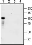
WB analysis of HEK-TrkC transfected cells (lanes 1 and 3) and HEK untransfected cells (lanes 2 and 4) lysates using GTX54858 TrkC antibody preincubated with or without immunogen peptide. Dilution : 1:400
 Panel B : Hoechst 33342 for nuclear staining (blue) Panel C : Merged images of Panel A and B Dilution : 1:50")
 appears in cell bodies. Note that the nerve fibers are not stained. Panel B : Nuclear staining (blue) was visualized using Hoechst 33342. Panel C : Merge of A and B. Dilution : 1:100")


WB analysis of HEK-TrkC transfected cells (lanes 1 and 3) and HEK untransfected cells (lanes 2 and 4) lysates using GTX54858 TrkC antibody preincubated with or without immunogen peptide. Dilution : 1:400
TrkC antibody
GTX54858
ApplicationsImmunoFluorescence, Western Blot, ImmunoCytoChemistry, ImmunoHistoChemistry, ImmunoHistoChemistry Frozen, ImmunoHistoChemistry Paraffin, Other Application
Product group Antibodies
ReactivityHuman, Mouse, Rat
TargetNtrk3
Overview
- SupplierGeneTex
- Product NameTrkC antibody
- Delivery Days Customer7
- ApplicationsImmunoFluorescence, Western Blot, ImmunoCytoChemistry, ImmunoHistoChemistry, ImmunoHistoChemistry Frozen, ImmunoHistoChemistry Paraffin, Other Application
- CertificationResearch Use Only
- ClonalityPolyclonal
- Concentration0.8 mg/ml
- ConjugateUnconjugated
- Gene ID29613
- Target nameNtrk3
- Target descriptionneurotrophic receptor tyrosine kinase 3
- Target synonymstrkC, NT-3 growth factor receptor, GP145-TrkC, neural receptor protein-tyrosine kinase (trkC), neurotrophic tyrosine kinase, receptor, type 3, trk-C, trkC tyrosine kinase
- HostRabbit
- IsotypeIgG
- Protein IDQ03351
- Protein NameNT-3 growth factor receptor
- Scientific Descriptiona tyrosine-protein kinase receptor for neurotrophin-3 [RGD, Feb 2006]
- ReactivityHuman, Mouse, Rat
- Storage Instruction-20°C or -80°C,2°C to 8°C
- UNSPSC41116161
References
- NT-3/TrkC Axis Contributes to the Perineural Invasion and the Poor Prognosis in Human Salivary Adenoid Cystic Carcinoma. Li H et al., 2019, J CancerRead this paper