

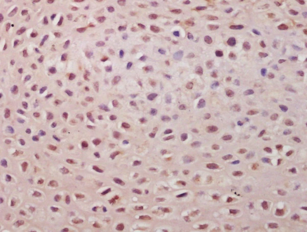
IHC-P analysis of mouse embryo tissue using GTX51813 TSPYL5 antibody. Dilution : 1:200
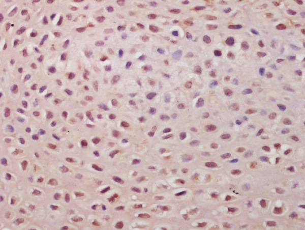
IHC-P analysis of mouse embryo tissue using GTX51813 TSPYL5 antibody. Dilution : 1:200
TSPYL5 antibody
GTX51813
ApplicationsImmunoHistoChemistry, ImmunoHistoChemistry Paraffin
Product group Antibodies
ReactivityHuman, Mouse
TargetTSPYL5
Overview
- SupplierGeneTex
- Product NameTSPYL5 antibody
- Delivery Days Customer9
- Application Supplier NoteIHC-P: 1:50-400. *Optimal dilutions/concentrations should be determined by the researcher.Not tested in other applications.
- ApplicationsImmunoHistoChemistry, ImmunoHistoChemistry Paraffin
- CertificationResearch Use Only
- ClonalityPolyclonal
- Concentration1 mg/ml
- ConjugateUnconjugated
- Gene ID85453
- Target nameTSPYL5
- Target descriptionTSPY like 5
- Target synonymstestis-specific Y-encoded-like protein 5, TSPY-like protein 5
- HostRabbit
- IsotypeIgG
- Protein IDQ86VY4
- Protein NameTestis-specific Y-encoded-like protein 5
- ReactivityHuman, Mouse
- Storage Instruction-20°C or -80°C,2°C to 8°C
- UNSPSC41116161
Datasheet
Related products

Product group Antibodies
Anti-TSPYL5 Antibody Picoband(r)A10648-2-CARRIER-FREE
ApplicationsFlow Cytometry, Western Blot, ELISA
ReactivityHuman
TargetTSPYL5
- SizePrice

Product group Antibodies
Anti-TSPYL5 AntibodyHPA031347
ApplicationsImmunoCytoChemistry, ImmunoHistoChemistry
ReactivityHuman
TargetTSPYL5
- SizePrice
Product group Antibodies

TSPYL5 Antibody (aa19-68)LS-C206212
ApplicationsWestern Blot
ReactivityHuman
TargetTSPYL5
- SizePrice
Product group Antibodies
TSPYL5 Polyclonal AntibodyBS-7703R
ApplicationsFlow Cytometry, ImmunoFluorescence, ELISA, ImmunoCytoChemistry, ImmunoHistoChemistry, ImmunoHistoChemistry Frozen, ImmunoHistoChemistry Paraffin
ReactivityEquine, Human, Mouse, Rabbit
TargetTSPYL5
- SizePrice