

Tyrosinase Antibody (Azide-free)
LS-C18404
ApplicationsImmunoFluorescence, Western Blot, ImmunoCytoChemistry, ImmunoHistoChemistry, ImmunoHistoChemistry Paraffin
Product group Antibodies
TargetTYR
Overview
- SupplierLifeSpan BioSciences
- Product NameTyrosinase Antibody (Azide-free)
- Delivery Days Customer23
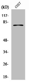
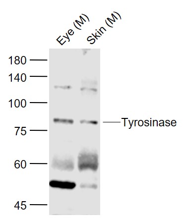
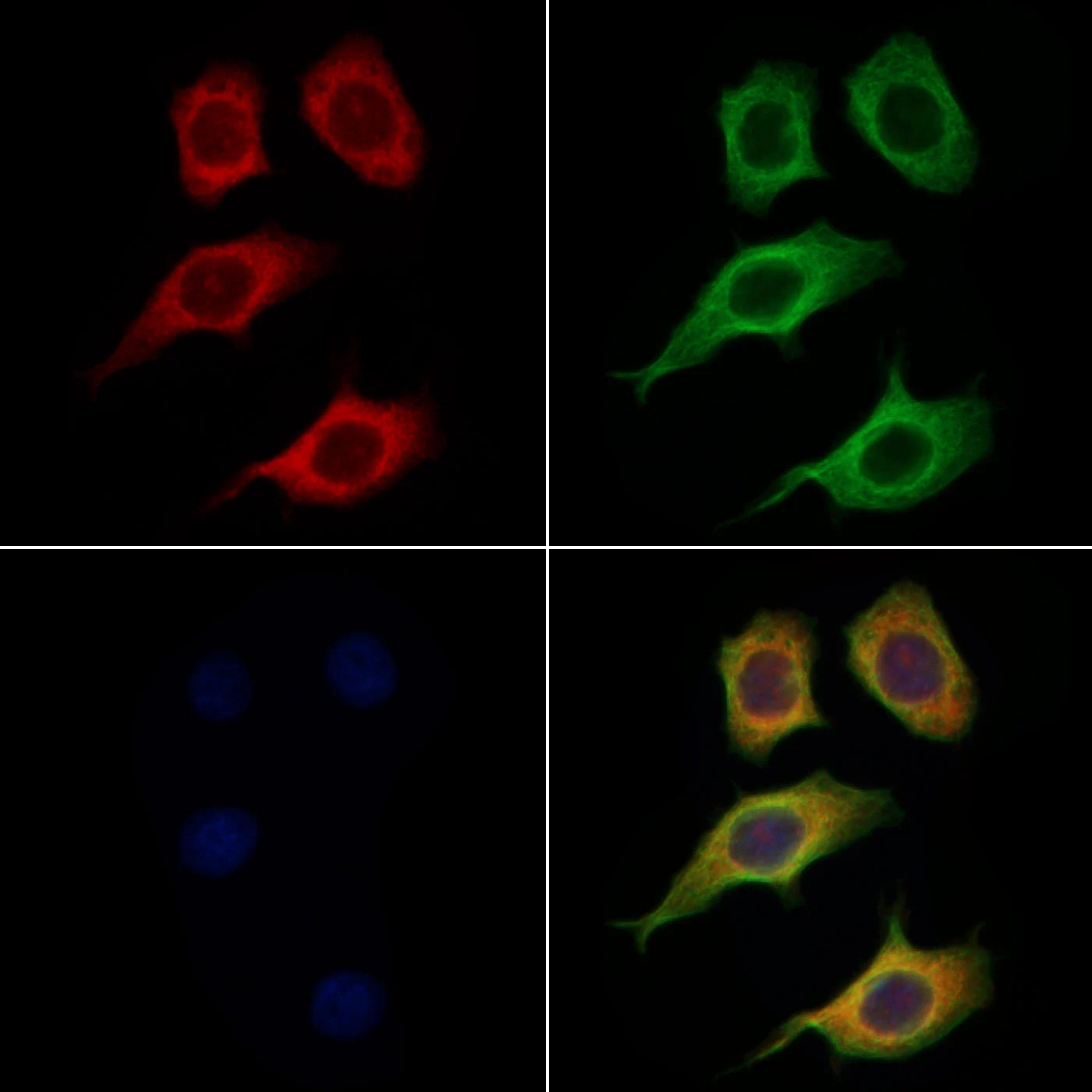
- Application Supplier NoteSuitable for use in Immunocytology, Immunofluorescence, Western Blot and Immunohistology. Western Blot: 1-2 ug/ml for 2 hours at RT. Immunohistology: 2-4 ug/ml for 30 minutes at RT (formalin/paraffin). Staining of formalin-fixed tissues requires boiling tissue sections in 1 mM EDTA, pH 8.0, for 10-20 min followed by cooling at RT for 20 min. Positive control: Melanoma cell lines (SK-MEL-13, SK-MEL-19, SK-MEL-30, SK-MEL-37), melanocytes in skin and Melanomas (1). The applications listed have been tested for the unmodified form of this product. Other forms have not been tested.. ICC, IF, IHC, IHC-P, WB (1 - 2 µg/ml) Suitable for use in Immunocytology, Immunofluorescence, Western Blot and Immunohistology. Western Blot: 1-2 ug/ml for 2 hours at RT. Immunohistology: 2-4 ug/ml for 30 minutes at RT (formalin/paraffin). Staining of formalin-fixed tissues requires boiling tissue sections in 1 mM EDTA, pH 8.0, for 10-20 min followed by cooling at RT for 20 min. Positive control: Melanoma cell lines (SK-MEL-13, SK-MEL-19, SK-MEL-30, SK-MEL-37), melanocytes in skin and Melanomas (1). The applications listed have been tested for the unmodified form of this product. Other forms have not been tested.
- ApplicationsImmunoFluorescence, Western Blot, ImmunoCytoChemistry, ImmunoHistoChemistry, ImmunoHistoChemistry Paraffin
- CertificationResearch Use Only
- ClonalityMonoclonal
- ConjugateUnconjugated
- Estimated Purity...
- Gene ID7299
- Target nameTYR
- Target descriptiontyrosinase
- Target synonymsATN; CMM8; LB24-AB; monophenol monooxygenase; OCA1; OCA1A; OCAIA; oculocutaneous albinism IA; SHEP3; SK29-AB; tumor rejection antigen AB; tyrosinase
- HostMouse
- IsotypeIgG2a
- Storage Instruction-20°C,2°C to 8°C
- UNSPSC12352203
Related products
Product group Antibodies
Anti-TYR Antibody144-01254
ApplicationsImmunoFluorescence, Western Blot, ImmunoHistoChemistry
TargetTYR
- SizePrice
Product group Antibodies
ApplicationsImmunoPrecipitation, Western Blot, ImmunoCytoChemistry, ImmunoHistoChemistry
TargetTYR
- SizePrice
Product group Antibodies
TYR AntibodyCSB-PA004353
ApplicationsWestern Blot, ELISA
ReactivityHuman, Monkey
TargetTYR
- SizePrice
Product group Antibodies

Tyrosinase AntibodyLS-C404376
ApplicationsWestern Blot, ELISA, ImmunoHistoChemistry
TargetTYR
- SizePrice
Product group Antibodies
References
Tyrosinase Polyclonal AntibodyBS-0819R
ApplicationsImmunoFluorescence, Western Blot, ELISA, ImmunoCytoChemistry, ImmunoHistoChemistry, ImmunoHistoChemistry Frozen, ImmunoHistoChemistry Paraffin
TargetTYR
- SizePrice

Product group Antibodies
Anti-TYR AntibodyHPA043241
ApplicationsImmunoCytoChemistry
ReactivityHuman
TargetTYR
- SizePrice
Product group Antibodies
Tyrosinase antibodyGTX04909
ApplicationsImmunoFluorescence, Western Blot, ImmunoCytoChemistry, ImmunoHistoChemistry, ImmunoHistoChemistry Paraffin
TargetTYR
- SizePrice

Product group Antibodies
Anti-Tyrosinase/TYR Antibody Picoband(r)A00326-3-CARRIER-FREE
ApplicationsWestern Blot, ELISA
TargetTYR
- SizePrice