

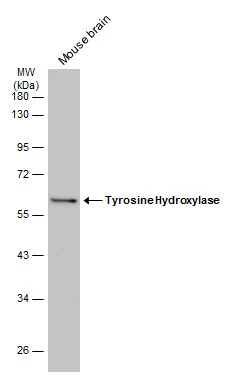
Mouse tissue extract (30 μg) was separated by 10% SDS-PAGE, and the membrane was blotted with Tyrosine Hydroxylase antibody [GT234] (GTX634481) diluted at 1:1000. The HRP-conjugated anti-mouse IgG antibody (GTX213111-01) was used to detect the primary antibody.
![Tyrosine Hydroxylase antibody [GT234] detects Tyrosine Hydroxylase protein in midbrain dopaminergic neurons by immunohistochemical analysis. Sample: Paraffin-embedded mouse brain. Green: Tyrosine Hydroxylase stained by Tyrosine Hydroxylase antibody [GT234] (GTX634481) diluted at 1:250. Blue: Fluoroshield with DAPI (GTX30920). Antigen Retrieval: Citrate buffer, pH 6.0, 15 min](https://www.genetex.com/upload/website/prouct_img/normal/GTX634481/GTX634481_43143_20181005_IHC-P-FL_M_w_23061202_340.webp "Tyrosine Hydroxylase antibody [GT234] detects Tyrosine Hydroxylase protein in midbrain dopaminergic neurons by immunohistochemical analysis. Sample: Paraffin-embedded mouse brain. Green: Tyrosine Hydroxylase stained by Tyrosine Hydroxylase antibody [GT234] (GTX634481) diluted at 1:250. Blue: Fluoroshield with DAPI (GTX30920). Antigen Retrieval: Citrate buffer, pH 6.0, 15 min")
![Rat tissue extract (50 μg) was separated by 7.5% SDS-PAGE, and the membrane was blotted with Tyrosine Hydroxylase antibody [GT234] (GTX634481) diluted at 1:1000. The HRP-conjugated anti-mouse IgG antibody (GTX213111-01) was used to detect the primary antibody.](https://www.genetex.com/upload/website/prouct_img/normal/GTX634481/GTX634481_43143_20180316_WB_R_brain_w_23061202_867.webp "Rat tissue extract (50 μg) was separated by 7.5% SDS-PAGE, and the membrane was blotted with Tyrosine Hydroxylase antibody [GT234] (GTX634481) diluted at 1:1000. The HRP-conjugated anti-mouse IgG antibody (GTX213111-01) was used to detect the primary antibody.")
![Tyrosine Hydroxylase antibody [GT234] detects Tyrosine Hydroxylase protein at cytoplasm of dopaminergic neurons by immunohistochemical analysis. Sample: Paraffin-embedded mouse midbrain. Green: Tyrosine Hydroxylase stained by Tyrosine Hydroxylase antibody [GT234] (GTX634481) diluted at 1:250. Blue: Fluoroshield with DAPI (GTX30920).
Antigen Retrieval: Citrate buffer, pH 6.0, 15 min](https://www.genetex.com/upload/website/prouct_img/normal/GTX634481/GTX634481_43143_20180713_IHC-P-FL_M_w_23061202_814.webp "Tyrosine Hydroxylase antibody [GT234] detects Tyrosine Hydroxylase protein at cytoplasm of dopaminergic neurons by immunohistochemical analysis. Sample: Paraffin-embedded mouse midbrain. Green: Tyrosine Hydroxylase stained by Tyrosine Hydroxylase antibody [GT234] (GTX634481) diluted at 1:250. Blue: Fluoroshield with DAPI (GTX30920).
Antigen Retrieval: Citrate buffer, pH 6.0, 15 min")
![Tyrosine Hydroxylase antibody [GT234] detects Tyrosine Hydroxylase protein by immunohistochemical analysis. Sample: Paraffin-embedded mouse eye. Green: Tyrosine Hydroxylase stained by Tyrosine Hydroxylase antibody [GT234] (GTX634481) diluted at 1:250. Red: beta Tubulin 3/ Tuj1 antibody (GTX130245) diluted at 1:250. Blue: Fluoroshield with DAPI (GTX30920). Antigen Retrieval: Citrate buffer, pH 6.0, 15 min](https://www.genetex.com/upload/website/prouct_img/normal/GTX634481/GTX634481_43143_20251031_IHC-P_M_25110619_154.webp "Tyrosine Hydroxylase antibody [GT234] detects Tyrosine Hydroxylase protein by immunohistochemical analysis. Sample: Paraffin-embedded mouse eye. Green: Tyrosine Hydroxylase stained by Tyrosine Hydroxylase antibody [GT234] (GTX634481) diluted at 1:250. Red: beta Tubulin 3/ Tuj1 antibody (GTX130245) diluted at 1:250. Blue: Fluoroshield with DAPI (GTX30920). Antigen Retrieval: Citrate buffer, pH 6.0, 15 min")
Mouse tissue extract (30 μg) was separated by 10% SDS-PAGE, and the membrane was blotted with Tyrosine Hydroxylase antibody [GT234] (GTX634481) diluted at 1:1000. The HRP-conjugated anti-mouse IgG antibody (GTX213111-01) was used to detect the primary antibody.
Tyrosine Hydroxylase antibody [GT234]
GTX634481
ApplicationsImmunoFluorescence, Western Blot, ImmunoCytoChemistry, ImmunoHistoChemistry, ImmunoHistoChemistry Paraffin
Product group Antibodies
ReactivityHuman, Mouse, Rat
TargetTH
Overview
- SupplierGeneTex
- Product NameTyrosine Hydroxylase antibody [GT234]
- Delivery Days Customer9
- Application Supplier NoteWB: 1:500-1:3000. IHC-P: 1:100-1:1000. *Optimal dilutions/concentrations should be determined by the researcher.Not tested in other applications.
- ApplicationsImmunoFluorescence, Western Blot, ImmunoCytoChemistry, ImmunoHistoChemistry, ImmunoHistoChemistry Paraffin
- CertificationResearch Use Only
- ClonalityMonoclonal
- Clone IDGT234
- Concentration1 mg/ml
- ConjugateUnconjugated
- Gene ID7054
- Target nameTH
- Target descriptiontyrosine hydroxylase
- Target synonymsDYT14, DYT5b, TYH, tyrosine 3-monooxygenase, dystonia 14, tyrosine 3-hydroxylase
- HostMouse
- IsotypeIgG1
- Protein IDP07101
- Protein NameTyrosine 3-monooxygenase
- Scientific DescriptionThe protein encoded by this gene is involved in the conversion of tyrosine to dopamine. It is the rate-limiting enzyme in the synthesis of catecholamines, hence plays a key role in the physiology of adrenergic neurons. Mutations in this gene have been associated with autosomal recessive Segawa syndrome. Alternatively spliced transcript variants encoding different isoforms have been noted for this gene. [provided by RefSeq, Jul 2008]
- ReactivityHuman, Mouse, Rat
- Storage Instruction-20°C or -80°C,2°C to 8°C
- UNSPSC12352203
References
- Feng X, Wang L, Zhou R, et al. Senescent immune cells accumulation promotes brown adipose tissue dysfunction during aging. Nat Commun. 2023,14(1):3208. doi: 10.1038/s41467-023-38842-6Read this paper
Datasheet
Related products
Product group Antibodies

ApplicationsImmunoFluorescence, Western Blot, ELISA, ImmunoCytoChemistry, ImmunoHistoChemistry
- SizePrice
Product group Antibodies
Anti-TH Antibody144-00028
ApplicationsImmunoFluorescence, Western Blot, ImmunoHistoChemistry
ReactivityHuman, Mouse, Rat
TargetTH
- SizePrice

Product group Antibodies
Anti-TH AntibodyAMAB91112
ApplicationsImmunoHistoChemistry
ReactivityHuman, Mouse, Rat
TargetTH
- SizePrice
Product group Antibodies
TH Polyclonal AntibodyCAC14884
ApplicationsWestern Blot, ELISA, ImmunoHistoChemistry
ReactivityMouse, Rat
TargetTH
- SizePrice
Product group Antibodies
References
ApplicationsImmunoFluorescence, Western Blot, ELISA, ImmunoCytoChemistry, ImmunoHistoChemistry, ImmunoHistoChemistry Frozen, ImmunoHistoChemistry Paraffin
ReactivityMouse, Rat
TargetTH
- SizePrice
Product group Antibodies
TH AntibodyCSB-PA004284
ApplicationsImmunoFluorescence, Western Blot, ELISA, ImmunoHistoChemistry
ReactivityHuman, Mouse, Rat
TargetTH
- SizePrice
![IHC-P analysis of human midbrain using GTX01922 Tyrosine Hydroxylase antibody [1B5]. Note cytoplasmic staining of catecholaminergic cells and their processes.](https://www.genetex.com/upload/website/prouct_img/normal/GTX01922/GTX01922_20200811_IHC-P_103_w_23053121_765.webp)
Product group Antibodies
ApplicationsImmunoHistoChemistry, ImmunoHistoChemistry Paraffin
ReactivityHuman, Mouse, Rat
TargetTH
- SizePrice
Product group Antibodies
ApplicationsImmunoFluorescence, ImmunoCytoChemistry
ReactivityHuman
TargetTH
- SizePrice