

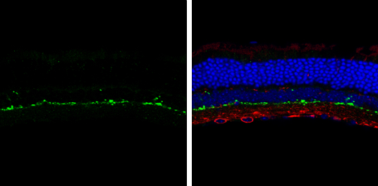
Tyrosine Hydroxylase antibody [N1C1] detects Tyrosine Hydroxylase protein by immunohistochemical analysis. Sample: Frozen sectioned adult mouse retina. Green: Tyrosine Hydroxylase protein stained by Tyrosine Hydroxylase antibody [N1C1] (GTX102424) diluted at 1:250. Red: beta Tubulin 3/ TUJ1, stained by beta Tubulin 3/ TUJ1 antibody [GT11710] (GTX631836) diluted at 1:250. Blue: Fluoroshield with DAPI (GTX30920).
![Whole cell extract (30 μg) was separated by 10% SDS-PAGE, and the membrane was blotted with Tyrosine Hydroxylase antibody [N1C1] (GTX102424) diluted at 1:1000. The HRP-conjugated anti-rabbit IgG antibody (GTX213110-01) was used to detect the primary antibody.](https://www.genetex.com/upload/website/prouct_img/normal/GTX102424/GTX102424_39974_20171020_WB_w_23060100_364.webp "Whole cell extract (30 μg) was separated by 10% SDS-PAGE, and the membrane was blotted with Tyrosine Hydroxylase antibody [N1C1] (GTX102424) diluted at 1:1000. The HRP-conjugated anti-rabbit IgG antibody (GTX213110-01) was used to detect the primary antibody.")
![Tyrosine Hydroxylase antibody [N1C1] detects Tyrosine Hydroxylase protein at cytoplasm by immunohistochemical analysis. Sample: Paraffin-embedded mouse brain. Tyrosine Hydroxylase stained by Tyrosine Hydroxylase antibody [N1C1] (GTX102424) diluted at 1:500. Antigen Retrieval: Citrate buffer, pH 6.0, 15 min](https://www.genetex.com/upload/website/prouct_img/normal/GTX102424/GTX102424_43810_20211005_IHC-P_M_1_w_23060100_180.webp "Tyrosine Hydroxylase antibody [N1C1] detects Tyrosine Hydroxylase protein at cytoplasm by immunohistochemical analysis. Sample: Paraffin-embedded mouse brain. Tyrosine Hydroxylase stained by Tyrosine Hydroxylase antibody [N1C1] (GTX102424) diluted at 1:500. Antigen Retrieval: Citrate buffer, pH 6.0, 15 min")
![Tyrosine Hydroxylase antibody [N1C1] detects Tyrosine Hydroxylase protein on zebrafish by whole mount immunohistochemical analysis. Sample: 2 days-post-fertilization zebrafish embryo. Tyrosine Hydroxylase antibody [N1C1] (GTX102424) dilution: 1:100.](https://www.genetex.com/upload/website/prouct_img/normal/GTX102424/GTX102424_39974_20160906_IHC-Wm_Z_1_w_23060100_501.webp "Tyrosine Hydroxylase antibody [N1C1] detects Tyrosine Hydroxylase protein on zebrafish by whole mount immunohistochemical analysis. Sample: 2 days-post-fertilization zebrafish embryo. Tyrosine Hydroxylase antibody [N1C1] (GTX102424) dilution: 1:100.")
![Tyrosine Hydroxylase antibody [N1C1] detects Tyrosine Hydroxylase protein at cytoplasm by immunohistochemical analysis. Sample: Paraffin-embedded mouse brain. Tyrosine Hydroxylase stained by Tyrosine Hydroxylase antibody [N1C1] (GTX102424) diluted at 1:1000. Antigen Retrieval: Citrate buffer, pH 6.0, 15 min](https://www.genetex.com/upload/website/prouct_img/normal/GTX102424/GTX102424_43810_20200313_IHC-P_M_w_23060100_460.webp "Tyrosine Hydroxylase antibody [N1C1] detects Tyrosine Hydroxylase protein at cytoplasm by immunohistochemical analysis. Sample: Paraffin-embedded mouse brain. Tyrosine Hydroxylase stained by Tyrosine Hydroxylase antibody [N1C1] (GTX102424) diluted at 1:1000. Antigen Retrieval: Citrate buffer, pH 6.0, 15 min")
![Tyrosine Hydroxylase antibody [N1C1] detects Tyrosine Hydroxylase protein at cell membrane and cytoplasm by immunohistochemical analysis. Sample: Paraffin-embedded mouse brain. Tyrosine Hydroxylase stained by Tyrosine Hydroxylase antibody [N1C1] (GTX102424) diluted at 1:500.
Antigen Retrieval: Citrate buffer, pH 6.0, 15 min](https://www.genetex.com/upload/website/prouct_img/normal/GTX102424/GTX102424_39974_20180403_IHC-P_M_1_w_23060100_416.webp "Tyrosine Hydroxylase antibody [N1C1] detects Tyrosine Hydroxylase protein at cell membrane and cytoplasm by immunohistochemical analysis. Sample: Paraffin-embedded mouse brain. Tyrosine Hydroxylase stained by Tyrosine Hydroxylase antibody [N1C1] (GTX102424) diluted at 1:500.
Antigen Retrieval: Citrate buffer, pH 6.0, 15 min")
![Tyrosine Hydroxylase antibody [N1C1] detects Tyrosine Hydroxylase protein at cytoplasm by immunofluorescent analysis. Sample: SK-N-SH cells were fixed in 4% paraformaldehyde at RT for 15 min. Green: Tyrosine Hydroxylase protein stained by Tyrosine Hydroxylase antibody [N1C1] (GTX102424) diluted at 1:200. Red: alpha Tubulin, a cytoskeleton marker, stained by alpha Tubulin antibody [GT114] (GTX628802) diluted at 1:1000. Blue: Hoechst 33342 staining.](https://www.genetex.com/upload/website/prouct_img/normal/GTX102424/GTX102424_39974_20150410_IFA_2_w_23060100_702.webp "Tyrosine Hydroxylase antibody [N1C1] detects Tyrosine Hydroxylase protein at cytoplasm by immunofluorescent analysis. Sample: SK-N-SH cells were fixed in 4% paraformaldehyde at RT for 15 min. Green: Tyrosine Hydroxylase protein stained by Tyrosine Hydroxylase antibody [N1C1] (GTX102424) diluted at 1:200. Red: alpha Tubulin, a cytoskeleton marker, stained by alpha Tubulin antibody [GT114] (GTX628802) diluted at 1:1000. Blue: Hoechst 33342 staining.")
![Tyrosine Hydroxylase antibody [N1C1] detects Tyrosine Hydroxylase protein at cytoplasm by immunohistochemical analysis. Sample: Paraffin-embedded mouse brain. Tyrosine Hydroxylase stained by Tyrosine Hydroxylase antibody [N1C1] (GTX102424) diluted at 1:500. Antigen Retrieval: Citrate buffer, pH 6.0, 15 min](https://www.genetex.com/upload/website/prouct_img/normal/GTX102424/GTX102424_43810_20211005_IHC-P_M_w_23060100_909.webp "Tyrosine Hydroxylase antibody [N1C1] detects Tyrosine Hydroxylase protein at cytoplasm by immunohistochemical analysis. Sample: Paraffin-embedded mouse brain. Tyrosine Hydroxylase stained by Tyrosine Hydroxylase antibody [N1C1] (GTX102424) diluted at 1:500. Antigen Retrieval: Citrate buffer, pH 6.0, 15 min")
![Various tissue extracts (50 μg) were separated by 10% SDS-PAGE, and the membrane was blotted with Tyrosine Hydroxylase antibody [N1C1] (GTX102424) diluted at 1:1000. The HRP-conjugated anti-rabbit IgG antibody (GTX213110-01) was used to detect the primary antibody.](https://www.genetex.com/upload/website/prouct_img/normal/GTX102424/GTX102424_43810_20191227_WB_M_R_w_23060100_505.webp "Various tissue extracts (50 μg) were separated by 10% SDS-PAGE, and the membrane was blotted with Tyrosine Hydroxylase antibody [N1C1] (GTX102424) diluted at 1:1000. The HRP-conjugated anti-rabbit IgG antibody (GTX213110-01) was used to detect the primary antibody.")
![Tyrosine Hydroxylase antibody [N1C1] detects Tyrosine Hydroxylase protein at cytoplasm by immunohistochemical analysis. Sample: Paraffin-embedded rat brain. Tyrosine Hydroxylase stained by Tyrosine Hydroxylase antibody [N1C1] (GTX102424) diluted at 1:1000. Antigen Retrieval: Citrate buffer, pH 6.0, 15 min](https://www.genetex.com/upload/website/prouct_img/normal/GTX102424/GTX102424_43810_20200313_IHC-P_R_w_23060100_995.webp "Tyrosine Hydroxylase antibody [N1C1] detects Tyrosine Hydroxylase protein at cytoplasm by immunohistochemical analysis. Sample: Paraffin-embedded rat brain. Tyrosine Hydroxylase stained by Tyrosine Hydroxylase antibody [N1C1] (GTX102424) diluted at 1:1000. Antigen Retrieval: Citrate buffer, pH 6.0, 15 min")
Tyrosine Hydroxylase antibody [N1C1] detects Tyrosine Hydroxylase protein by immunohistochemical analysis. Sample: Frozen sectioned adult mouse retina. Green: Tyrosine Hydroxylase protein stained by Tyrosine Hydroxylase antibody [N1C1] (GTX102424) diluted at 1:250. Red: beta Tubulin 3/ TUJ1, stained by beta Tubulin 3/ TUJ1 antibody [GT11710] (GTX631836) diluted at 1:250. Blue: Fluoroshield with DAPI (GTX30920).
Tyrosine Hydroxylase antibody [N1C1]
GTX102424
ApplicationsImmunoFluorescence, Western Blot, ImmunoCytoChemistry, ImmunoHistoChemistry, ImmunoHistoChemistry Frozen, ImmunoHistoChemistry Paraffin
Product group Antibodies
ReactivityHuman, Mouse, Rat, Zebra Fish
TargetTH
Overview
- SupplierGeneTex
- Product NameTyrosine Hydroxylase antibody [N1C1]
- Delivery Days Customer9
- Application Supplier NoteWB: 1:500-1:3000. ICC/IF: 1:100-1:1000. IHC-P: 1:100-1:1000. IHC-Fr: 1:100-1:1000. IHC-Wm: 1:100-1:500. *Optimal dilutions/concentrations should be determined by the researcher.Not tested in other applications.
- ApplicationsImmunoFluorescence, Western Blot, ImmunoCytoChemistry, ImmunoHistoChemistry, ImmunoHistoChemistry Frozen, ImmunoHistoChemistry Paraffin
- CertificationResearch Use Only
- ClonalityPolyclonal
- Concentration0.57 mg/ml
- ConjugateUnconjugated
- Gene ID7054
- Target nameTH
- Target descriptiontyrosine hydroxylase
- Target synonymsDYT14, DYT5b, TYH, tyrosine 3-monooxygenase, dystonia 14, tyrosine 3-hydroxylase
- HostRabbit
- IsotypeIgG
- Protein IDP07101
- Protein NameTyrosine 3-monooxygenase
- Scientific DescriptionThe protein encoded by this gene is involved in the conversion of tyrosine to dopamine. It is the rate-limiting enzyme in the synthesis of catecholamines, hence plays a key role in the physiology of adrenergic neurons. Mutations in this gene have been associated with autosomal recessive Segawa syndrome. Alternatively spliced transcript variants encoding different isoforms have been noted for this gene. [provided by RefSeq]
- ReactivityHuman, Mouse, Rat, Zebra Fish
- Storage Instruction-20°C or -80°C,2°C to 8°C
- UNSPSC41116161
Datasheet
Related products
Product group Antibodies
TH AntibodyCSB-PA004284
ApplicationsImmunoFluorescence, Western Blot, ELISA, ImmunoHistoChemistry
ReactivityHuman, Mouse, Rat
TargetTH
- SizePrice
Product group Antibodies
ApplicationsFlow Cytometry, ImmunoFluorescence, Western Blot, ImmunoCytoChemistry, ImmunoHistoChemistry
ReactivityHuman, Mouse, Rat
- SizePrice
Product group Antibodies

ApplicationsImmunoFluorescence, Western Blot, ELISA, ImmunoCytoChemistry, ImmunoHistoChemistry
- SizePrice

Product group Antibodies
Anti-TH AntibodyAMAB91112
ApplicationsImmunoHistoChemistry
ReactivityHuman, Mouse, Rat
TargetTH
- SizePrice
Product group Antibodies

TH / Tyrosine Hydroxylase AntibodyLS-C832610
ApplicationsELISA, ImmunoHistoChemistry
ReactivityHuman
TargetTH
- SizePrice
Product group Antibodies

ApplicationsELISA, ImmunoHistoChemistry
ReactivityCanine, Human, Rat
TargetTH
- SizePrice
Product group Antibodies
TH Polyclonal AntibodyCAC14884
ApplicationsWestern Blot, ELISA, ImmunoHistoChemistry
ReactivityMouse, Rat
TargetTH
- SizePrice

Product group Antibodies
Anti-Tyrosine Hydroxylase/TH Antibody Picoband(r)PB9449-CARRIER-FREE
ApplicationsImmunoFluorescence, Western Blot, ImmunoHistoChemistry
ReactivityHamster, Mouse, Rat
TargetTH
- SizePrice
![Whole cell extract (30 μg) was separated by 10% SDS-PAGE, and the membrane was blotted with Tyrosine Hydroxylase antibody [HL1762] (GTX637412) diluted at 1:1000. The HRP-conjugated anti-rabbit IgG antibody (GTX213110-01) was used to detect the primary antibody.](https://www.genetex.com/upload/website/prouct_img/normal/GTX637412/GTX637412_T-44795_20220908_WB_R_22091323_117.webp)
Product group Antibodies
ApplicationsWestern Blot, ImmunoHistoChemistry, ImmunoHistoChemistry Frozen, ImmunoHistoChemistry Paraffin
ReactivityHuman, Mouse, Rat
TargetTH
- SizePrice