

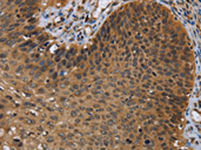
The image on the left is immunohistochemistry of paraffin-embedded Human cervical cancer tissue using CSB-PA049538(UHRF2 Antibody) at dilution 1/20, on the right is treated with synthetic peptide. (Original magnification: x200)
 at dilution 1/20, on the right is treated with synthetic peptide. (Original magnification: x200)")
The image on the left is immunohistochemistry of paraffin-embedded Human cervical cancer tissue using CSB-PA049538(UHRF2 Antibody) at dilution 1/20, on the right is treated with synthetic peptide. (Original magnification: x200)
UHRF2 Antibody
CSB-PA049538
ApplicationsELISA, ImmunoHistoChemistry
Product group Antibodies
ReactivityHuman, Mouse
TargetUHRF2
Overview
- SupplierCusabio
- Product NameUHRF2 Antibody
- Delivery Days Customer20
- ApplicationsELISA, ImmunoHistoChemistry
- CertificationResearch Use Only
- ClonalityPolyclonal
- ConjugateUnconjugated
- Gene ID115426
- Target nameUHRF2
- Target descriptionubiquitin like with PHD and ring finger domains 2
- Target synonymsNIRF, RNF107, TDRD23, URF2, E3 ubiquitin-protein ligase UHRF2, Np95-like ring finger protein, RING finger protein 107, RING-type E3 ubiquitin transferase UHRF2, np95/ICBP90-like RING finger protein, nuclear protein 97, nuclear zinc finger protein NP97, ubiquitin-like PHD and RING finger domain-containing protein 2, ubiquitin-like with PHD and ring finger domains 2, E3 ubiquitin protein ligase, ubiquitin-like, containing PHD and RING finger domains, 2, ubiquitin-like-containing PHD and RING finger domains protein 2
- HostRabbit
- IsotypeIgG
- Protein IDQ96PU4
- Protein NameE3 ubiquitin-protein ligase UHRF2
- Scientific DescriptionThis gene encodes a nuclear protein which is involved in cell-cycle regulation. The encoded protein is a ubiquitin-ligase capable of ubiquinating PCNP (PEST-containing nuclear protein), and together they may play a role in tumorigenesis. The encoded protein contains an NIRF_N domain, a PHD finger, a set- and ring-associated (SRA) domain, and a RING finger domain and several of these domains have been shown to be essential for the regulation of cell proliferation. This protein may also have a role in intranuclear degradation of polyglutamine aggregates. Alternative splicing results in multiple transcript variants some of which are non-protein coding.
- ReactivityHuman, Mouse
- Storage Instruction-20°C or -80°C
- UNSPSC41116161
Related products
Product group Antibodies

Anti-UHRF2 [RAB-C515]Ab01915-1.1
ApplicationsFlow Cytometry, ImmunoFluorescence
ReactivityHuman
TargetUHRF2
- SizePrice
Product group Antibodies
Anti-UHRF2 Antibody101-11894
ApplicationsImmunoFluorescence, Western Blot, ELISA
TargetUHRF2
- SizePrice
Product group Antibodies
NIRF Polyclonal AntibodyBS-6389R
ApplicationsImmunoFluorescence, ELISA, ImmunoCytoChemistry, ImmunoHistoChemistry, ImmunoHistoChemistry Frozen, ImmunoHistoChemistry Paraffin
ReactivityBovine, Canine, Human, Mouse, Rat
TargetUHRF2
- SizePrice
Product group Antibodies

NIRF / UHRF2 AntibodyLS-C403367
ApplicationsELISA, ImmunoHistoChemistry
ReactivityHuman, Mouse
TargetUHRF2
- SizePrice

Product group Antibodies
Anti-UHRF2 AntibodyHPA026633
ApplicationsImmunoCytoChemistry
ReactivityHuman
TargetUHRF2
- SizePrice

Product group Antibodies
Anti-NIRF/UHRF2 Antibody Picoband(r)PB9906-CARRIER-FREE
ApplicationsWestern Blot, ImmunoHistoChemistry
ReactivityHuman, Mouse, Rat
TargetUHRF2
- SizePrice