

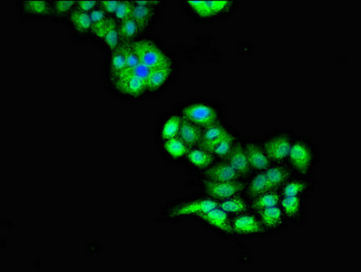
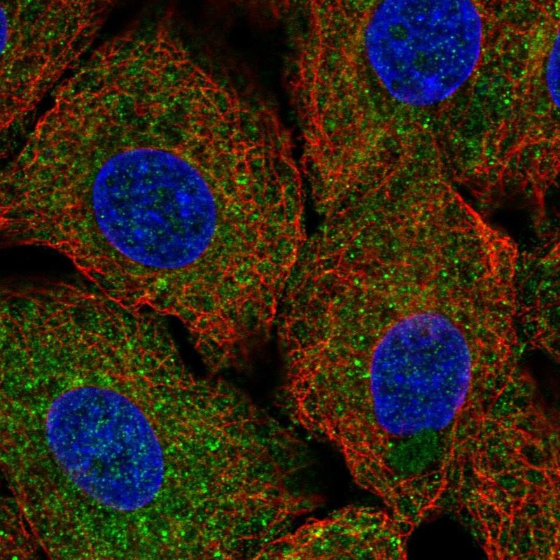
Immunofluorescent analysis of PC-3 cells using CSB-PA025612LA01HU at dilution of 1:100 and Alexa Fluor 488-congugated AffiniPure Goat Anti-Rabbit IgG(H+L)
Immunofluorescent analysis of PC-3 cells using CSB-PA025612LA01HU at dilution of 1:100 and Alexa Fluor 488-congugated AffiniPure Goat Anti-Rabbit IgG(H+L)
ULK1 Antibody
CSB-PA025612LA01HU
ApplicationsImmunoFluorescence, ELISA
Product group Antibodies
ReactivityHuman
TargetULK1
Overview
- SupplierCusabio
- Product NameULK1 Antibody
- Delivery Days Customer20
- ApplicationsImmunoFluorescence, ELISA
- CertificationResearch Use Only
- ClonalityPolyclonal
- ConjugateUnconjugated
- Gene ID8408
- Target nameULK1
- Target descriptionunc-51 like autophagy activating kinase 1
- Target synonymsATG1, ATG1A, UNC51, Unc51.1, hATG1, serine/threonine-protein kinase ULK1, ATG1 autophagy related 1 homolog, autophagy-related protein 1 homolog
- HostRabbit
- IsotypeIgG
- Protein IDO75385
- Protein NameSerine/threonine-protein kinase ULK1
- Scientific DescriptionSerine/threonine-protein kinase involved in autophagy in response to starvation. Acts upstream of phosphatidylinositol 3-kinase PIK3C3 to regulate the formation of autophagophores, the precursors of autophagosomes. Part of regulatory feedback loops in autophagy: acts both as a downstream effector and negative regulator of mammalian target of rapamycin complex 1 (mTORC1) via interaction with RPTOR. Activated via phosphorylation by AMPK and also acts as a regulator of AMPK by mediating phosphorylation of AMPK subunits PRKAA1, PRKAB2 and PRKAG1, leading to negatively regulate AMPK activity. May phosphorylate ATG13/KIAA0652 and RPTOR; however such data need additional evidences. Plays a role early in neuronal differentiation and is required for granule cell axon formation. May also phosphorylate SESN2 and SQSTM1 to regulate autophagy (PubMed:25040165).
- ReactivityHuman
- Storage Instruction-20°C or -80°C
- UNSPSC41116161
Related products
Product group Antibodies
Anti-ULK1 AntibodyA10448
ApplicationsImmunoFluorescence, Western Blot, ImmunoCytoChemistry
ReactivityHuman, Mouse, Rat
- SizePrice
Product group Antibodies
Anti-ULk1 Antibody144-66638
ApplicationsImmunoFluorescence, Western Blot, ImmunoHistoChemistry
ReactivityHuman, Mouse, Rat
TargetULK1
- SizePrice

Product group Antibodies
Anti-ULK1 Antibody Picoband(r)A00584-1-CARRIER-FREE
ApplicationsFlow Cytometry, Western Blot, ImmunoHistoChemistry
ReactivityHuman, Mouse, Rat
TargetULK1
- SizePrice
Product group Antibodies
References
ATG1/ULK1 Polyclonal AntibodyBS-3602R
ApplicationsFlow Cytometry, ImmunoFluorescence, Western Blot, ELISA, ImmunoCytoChemistry, ImmunoHistoChemistry, ImmunoHistoChemistry Frozen, ImmunoHistoChemistry Paraffin
ReactivityBovine, Chicken, Equine, Human, Mouse, Porcine, Rabbit, Rat
TargetULK1
- SizePrice
Product group Antibodies

ULK1 Antibody (aa400-499)LS-C410064
ApplicationsImmunoFluorescence, Western Blot
ReactivityHuman
TargetULK1
- SizePrice
Product group Antibodies
ULK1 (phospho Ser758) antibodyGTX132654
ApplicationsWestern Blot
ReactivityHuman
TargetULK1
- SizePrice
Product group Antibodies
Anti-ULK1 AntibodyHPA063990
ApplicationsImmunoCytoChemistry
ReactivityHuman
TargetULK1
- SizePrice
Product group Antibodies
Anti-ULK1/ULK2 AntibodyCAB18685
ApplicationsWestern Blot, ELISA
ReactivityHuman
TargetULK1
- SizePrice
Product group Antibodies
anti-Phospho-ULk1 (Autophagy-related Protein 1 Homolog, ATG1) (Ser757), Rabbit Monoclonal (RM488)REV-31-1380-00
ApplicationsWestern Blot
ReactivityHuman, Mouse, Rat
TargetULK1
- SizePrice