


ULK2 antibody [C2C3], C-term detects ULK2 protein at cytoplasm by immunohistochemical analysis. Sample: Paraffin-embedded rat brain. ULK2 stained by ULK2 antibody [C2C3], C-term (GTX111476) diluted at 1:500. Antigen Retrieval: Citrate buffer, pH 6.0, 15 min
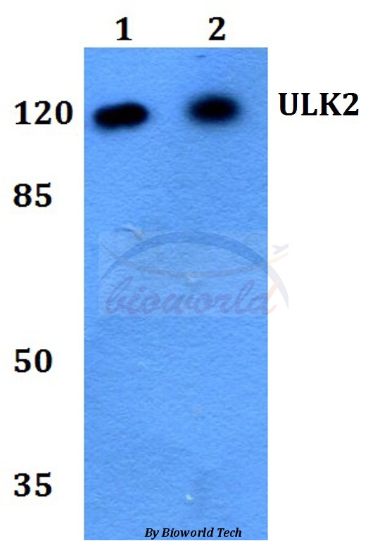
![Non-transfected (–) and transfected (+) 293T whole cell extracts (30 μg) were separated by 7.5% SDS-PAGE, and the membrane was blotted with ULK2 antibody [C2C3], C-term (GTX111476) diluted at 1:1000. The HRP-conjugated anti-rabbit IgG antibody (GTX213110-01) was used to detect the primary antibody.](https://www.genetex.com/upload/website/prouct_img/normal/GTX111476/GTX111476_44258_20221216_WB_shRNA_watermark_22122018_879.webp "Non-transfected (–) and transfected (+) 293T whole cell extracts (30 μg) were separated by 7.5% SDS-PAGE, and the membrane was blotted with ULK2 antibody [C2C3], C-term (GTX111476) diluted at 1:1000. The HRP-conjugated anti-rabbit IgG antibody (GTX213110-01) was used to detect the primary antibody.")
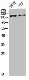
![Various whole cell extracts (30 μg) were separated by 7.5% SDS-PAGE, and the membrane was blotted with ULK2 antibody [C2C3], C-term (GTX111476) diluted at 1:500. The HRP-conjugated anti-rabbit IgG antibody (GTX213110-01) was used to detect the primary antibody.](https://www.genetex.com/upload/website/prouct_img/normal/GTX111476/GTX111476_44258_20210326_WB_w_23060500_257.webp "Various whole cell extracts (30 μg) were separated by 7.5% SDS-PAGE, and the membrane was blotted with ULK2 antibody [C2C3], C-term (GTX111476) diluted at 1:500. The HRP-conjugated anti-rabbit IgG antibody (GTX213110-01) was used to detect the primary antibody.")
![ULK2 antibody [C2C3], C-term detects ULK2 protein by western blot analysis. Whole cell extracts (30 μg) was separated by 5% SDS-PAGE, and the membrane was blotted with ULK2 antibody [C2C3], C-term (GTX111476) at a dilution of 1:1000. The HRP-conjugated anti-rabbit IgG antibody (GTX213110-01) was used to detect the primary antibody.](https://www.genetex.com/upload/website/prouct_img/normal/GTX111476/GTX111476_41703_20150529_WB_w_23060500_681.webp "ULK2 antibody [C2C3], C-term detects ULK2 protein by western blot analysis. Whole cell extracts (30 μg) was separated by 5% SDS-PAGE, and the membrane was blotted with ULK2 antibody [C2C3], C-term (GTX111476) at a dilution of 1:1000. The HRP-conjugated anti-rabbit IgG antibody (GTX213110-01) was used to detect the primary antibody.")
![ULK2 antibody [C2C3], C-term detects ULK2 protein at cytoplasm and nucleus by immunofluorescent analysis. Sample: HeLa cells were fixed in ice-cold MeOH for 5 min. Green: ULK2 stained by ULK2 antibody [C2C3], C-term (GTX111476) diluted at 1:500. Blue: Hoechst 33342 staining. Scale bar= 10 μm.](https://www.genetex.com/upload/website/prouct_img/normal/GTX111476/GTX111476_41703_20190116_ICC_IF_w_23060500_694.webp "ULK2 antibody [C2C3], C-term detects ULK2 protein at cytoplasm and nucleus by immunofluorescent analysis. Sample: HeLa cells were fixed in ice-cold MeOH for 5 min. Green: ULK2 stained by ULK2 antibody [C2C3], C-term (GTX111476) diluted at 1:500. Blue: Hoechst 33342 staining. Scale bar= 10 μm.")
 antibody at 1:500 dilution.
Antigen Retrieval: Trilogy? (EDTA based, pH 8.0) buffer, 15min")
![Various whole cell extracts (30 μg) were separated by 7.5% SDS-PAGE, and the membrane was blotted with ULK2 antibody [C2C3], C-term (GTX111476) diluted at 1:500. The HRP-conjugated anti-rabbit IgG antibody (GTX213110-01) was used to detect the primary antibody.](https://www.genetex.com/upload/website/prouct_img/normal/GTX111476/GTX111476_41703_20190201_WB_M_w_23060500_644.webp "Various whole cell extracts (30 μg) were separated by 7.5% SDS-PAGE, and the membrane was blotted with ULK2 antibody [C2C3], C-term (GTX111476) diluted at 1:500. The HRP-conjugated anti-rabbit IgG antibody (GTX213110-01) was used to detect the primary antibody.")
 antibody at 1:200 dilution.")
![Various whole cell extracts (30 μg) were separated by 7.5% SDS-PAGE, and the membrane was blotted with ULK2 antibody [C2C3], C-term (GTX111476) diluted at 1:500. The HRP-conjugated anti-rabbit IgG antibody (GTX213110-01) was used to detect the primary antibody.Corresponding RNA expression data for the same cell lines are based on Human Protein Atlas program.](https://www.genetex.com/upload/website/prouct_img/normal/GTX111476/GTX111476_44727_20240830_WB_TPM_watermark_24090423_576.webp "Various whole cell extracts (30 μg) were separated by 7.5% SDS-PAGE, and the membrane was blotted with ULK2 antibody [C2C3], C-term (GTX111476) diluted at 1:500. The HRP-conjugated anti-rabbit IgG antibody (GTX213110-01) was used to detect the primary antibody.Corresponding RNA expression data for the same cell lines are based on Human Protein Atlas program.")
ULK2 antibody [C2C3], C-term detects ULK2 protein at cytoplasm by immunohistochemical analysis. Sample: Paraffin-embedded rat brain. ULK2 stained by ULK2 antibody [C2C3], C-term (GTX111476) diluted at 1:500. Antigen Retrieval: Citrate buffer, pH 6.0, 15 min
ULK2 antibody [C2C3], C-term
GTX111476
ApplicationsImmunoFluorescence, Western Blot, ImmunoCytoChemistry, ImmunoHistoChemistry, ImmunoHistoChemistry Paraffin
Product group Antibodies
ReactivityHuman, Mouse, Rat
TargetULK2
Overview
- SupplierGeneTex
- Product NameULK2 antibody [C2C3], C-term
- Delivery Days Customer9
- Application Supplier NoteWB: 1:500-1:3000. ICC/IF: 1:100-1:1000. IHC-P: 1:100-1:1000. *Optimal dilutions/concentrations should be determined by the researcher.Not tested in other applications.
- ApplicationsImmunoFluorescence, Western Blot, ImmunoCytoChemistry, ImmunoHistoChemistry, ImmunoHistoChemistry Paraffin
- CertificationResearch Use Only
- ClonalityPolyclonal
- Concentration0.21 mg/ml
- ConjugateUnconjugated
- Gene ID9706
- Target nameULK2
- Target descriptionunc-51 like autophagy activating kinase 2
- Target synonymsATG1B, Unc51.2, serine/threonine-protein kinase ULK2
- HostRabbit
- IsotypeIgG
- Protein IDQ8IYT8
- Protein NameSerine/threonine-protein kinase ULK2
- Scientific DescriptionThis gene encodes a protein that is similar to a serine/threonine kinase in C. elegans which is involved in axonal elongation. The structure of this protein is similar to the C. elegans protein in that both proteins have an N-terminal kinase domain, a central proline/serine rich (PS) domain, and a C-terminal (C) domain. The gene is located within the Smith-Magenis syndrome region on chromosome 17. Alternatively spliced transcript variants encoding the same protein have been identified. [provided by RefSeq]
- ReactivityHuman, Mouse, Rat
- Storage Instruction-20°C or -80°C,2°C to 8°C
- UNSPSC41116161
Datasheet
Related products
Product group Antibodies
ULK2 AntibodyCSB-PA461940
ApplicationsWestern Blot, ELISA
ReactivityHuman, Mouse, Rat
TargetULK2
- SizePrice

Product group Antibodies
Anti-ULK2 AntibodyA05219
ApplicationsWestern Blot, ELISA
ReactivityHuman, Mouse, Rat
TargetULK2
- SizePrice
Product group Antibodies

ULK2 AntibodyLS-C750213
ApplicationsWestern Blot
ReactivityHuman, Rat
TargetULK2
- SizePrice
Product group Antibodies
ULK2 Polyclonal AntibodyCAC15441
ApplicationsWestern Blot, ELISA, ImmunoHistoChemistry
TargetULK2
- SizePrice
![Whole cell extract (30 μg) was separated by 7.5% SDS-PAGE, and the membrane was blotted with ULK2 antibody [HL2206] (GTX638210) diluted at 1:1000. The HRP-conjugated anti-rabbit IgG antibody (GTX213110-01) was used to detect the primary antibody.](https://www.genetex.com/upload/website/prouct_img/normal/GTX638210/GTX638210_T-44942_20230210_WB_R_23021401_368.webp)
Product group Antibodies
ULK2 antibody [HL2206]GTX638210
ApplicationsImmunoFluorescence, Western Blot, ImmunoCytoChemistry
ReactivityHuman, Rat
TargetULK2
- SizePrice