

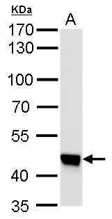
UQCRC1 antibody [GT139] detects UQCRC1 protein by western blot analysis. A. 50 μg mouse brain lysate/extract 7.5 % SDS-PAGE UQCRC1 antibody [GT139] (GTX630413) dilution: 1:1000
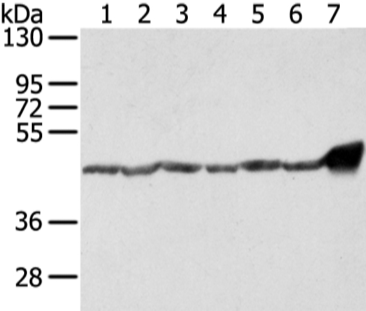
![UQCRC1 antibody [GT139] detects UQCRC1 protein by western blot analysis. A. 50 μg rat brain lysate/extract 10 % SDS-PAGE UQCRC1 antibody [GT139] (GTX630413) dilution: 1:1000](https://www.genetex.com/upload/website/prouct_img/normal/GTX630413/GTX630413_41540_WB_R_brain_w_23061202_693.webp "UQCRC1 antibody [GT139] detects UQCRC1 protein by western blot analysis. A. 50 μg rat brain lysate/extract 10 % SDS-PAGE UQCRC1 antibody [GT139] (GTX630413) dilution: 1:1000")
![UQCRC1 antibody [GT139] detects UQCRC1 protein at mitochondria by immunofluorescent analysis. Sample: HeLa cells were fixed in 4% paraformaldehyde at room temperature. Green: UQCRC1 protein stained by UQCRC1 antibody [GT139] (GTX630413) diluted at 1:100. Blue: Hoechst 33342 staining. Scale bar = 10 μm.](https://www.genetex.com/upload/website/prouct_img/normal/GTX630413/GTX630413_41540_20150821_IFA_w_23061202_955.webp "UQCRC1 antibody [GT139] detects UQCRC1 protein at mitochondria by immunofluorescent analysis. Sample: HeLa cells were fixed in 4% paraformaldehyde at room temperature. Green: UQCRC1 protein stained by UQCRC1 antibody [GT139] (GTX630413) diluted at 1:100. Blue: Hoechst 33342 staining. Scale bar = 10 μm.")
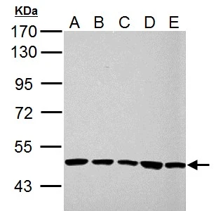
![UQCRC1 antibody [GT139] detects UQCRC1 protein by western blot analysis. A. 30 μg 293T whole cell lysate/extract B. 30 μg A431 whole cell lysate/extract C. 30 μg HeLa whole cell lysate/extract D. 30 μg HepG2 whole cell lysate/extract 10 % SDS-PAGE UQCRC1 antibody [GT139] (GTX630413) dilution: 1:1000](https://www.genetex.com/upload/website/prouct_img/normal/GTX630413/GTX630413_41540_WB_w_23061202_132.webp "UQCRC1 antibody [GT139] detects UQCRC1 protein by western blot analysis. A. 30 μg 293T whole cell lysate/extract B. 30 μg A431 whole cell lysate/extract C. 30 μg HeLa whole cell lysate/extract D. 30 μg HepG2 whole cell lysate/extract 10 % SDS-PAGE UQCRC1 antibody [GT139] (GTX630413) dilution: 1:1000")
![Non-transfected (–) and transfected (+) 293T whole cell extracts (30 μg) were separated by 10% SDS-PAGE, and the membrane was blotted with UQCRC1 antibody [GT139] (GTX630413) diluted at 1:500.](https://www.genetex.com/upload/website/prouct_img/normal/GTX630413/GTX630413_41540_20161103_WB_shRNA_watermark_w_23061202_995.webp "Non-transfected (–) and transfected (+) 293T whole cell extracts (30 μg) were separated by 10% SDS-PAGE, and the membrane was blotted with UQCRC1 antibody [GT139] (GTX630413) diluted at 1:500.")
UQCRC1 antibody [GT139] detects UQCRC1 protein by western blot analysis. A. 50 μg mouse brain lysate/extract 7.5 % SDS-PAGE UQCRC1 antibody [GT139] (GTX630413) dilution: 1:1000
UQCRC1 antibody [GT139]
GTX630413
ApplicationsImmunoFluorescence, Western Blot, ImmunoCytoChemistry
Product group Antibodies
ReactivityHuman, Mouse, Rat
TargetUQCRC1
Overview
- SupplierGeneTex
- Product NameUQCRC1 antibody [GT139]
- Delivery Days Customer9
- Application Supplier NoteWB: 1:500-1:3000. ICC/IF: 1:100-1:1000. *Optimal dilutions/concentrations should be determined by the researcher.Not tested in other applications.
- ApplicationsImmunoFluorescence, Western Blot, ImmunoCytoChemistry
- CertificationResearch Use Only
- ClonalityMonoclonal
- Clone IDGT139
- Concentration1 mg/ml
- ConjugateUnconjugated
- Gene ID7384
- Target nameUQCRC1
- Target descriptionubiquinol-cytochrome c reductase core protein 1
- Target synonymsD3S3191, PKNPY, QCR1, UQCR1, cytochrome b-c1 complex subunit 1, mitochondrial, complex III subunit 1, core protein I, ubiquinol-cytochrome c reductase core protein I, ubiquinol-cytochrome-c reductase complex core protein 1
- HostMouse
- IsotypeIgG1
- Protein IDP31930
- Protein NameCytochrome b-c1 complex subunit 1, mitochondrial
- Scientific DescriptionThis is a component of the ubiquinol-cytochrome c reductase complex (complex III or cytochrome b-c1 complex), which is part of the mitochondrial respiratory chain. This protein may mediate formation of the complex between cytochromes c and c1.
- ReactivityHuman, Mouse, Rat
- Storage Instruction-20°C or -80°C,2°C to 8°C
- UNSPSC41116161
Datasheet
Related products
Product group Antibodies
Anti-UQCRC1 AntibodyA37382
ApplicationsWestern Blot, ImmunoHistoChemistry
ReactivityHuman
- SizePrice
Product group Antibodies
Anti-UQCRC1 Antibody144-03339
ApplicationsWestern Blot, ImmunoHistoChemistry
ReactivityHuman, Monkey, Mouse, Rat
TargetUQCRC1
- SizePrice
Product group Antibodies
UQCRC1 AntibodyCSB-PA025667ESR2HU
ApplicationsWestern Blot, ELISA, ImmunoHistoChemistry
ReactivityHuman, Mouse
TargetUQCRC1
- SizePrice
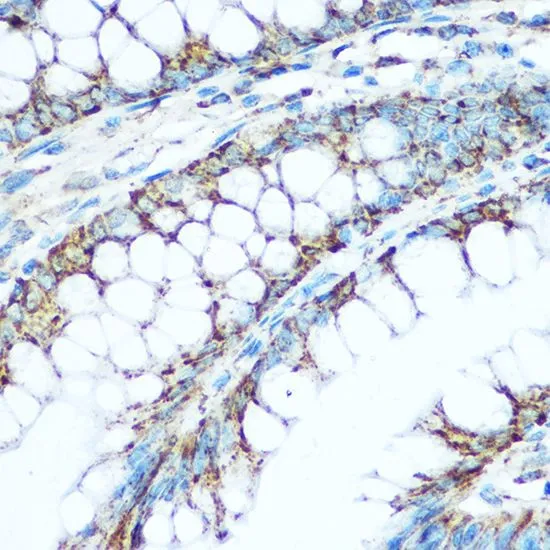
![UQCRC1 antibody [N1N3] detects UQCRC1 protein at mitochondria on mouse liver by immunohistochemical analysis. Sample: Paraffin-embedded mouse liver. UQCRC1 antibody [N1N3] (GTX101862) diluted at 1:500.
Antigen Retrieval: Trilogy? (EDTA based, pH 8.0) buffer, 15min](https://www.genetex.com/upload/website/prouct_img/normal/GTX101862/GTX101862_39568_20141205_IHC_M_w_23060100_525.webp)
Product group Antibodies
UQCRC1 antibody [N1N3]GTX101862
ApplicationsWestern Blot, ImmunoHistoChemistry, ImmunoHistoChemistry Paraffin
ReactivityHuman, Mouse
TargetUQCRC1
- SizePrice
Product group Antibodies
UQCRC1 antibodyGTX101896
ApplicationsImmunoFluorescence, Western Blot, ImmunoCytoChemistry
ReactivityHuman
TargetUQCRC1
- SizePrice
Product group Antibodies

UQCRC1 AntibodyLS-C332502
ApplicationsWestern Blot, ImmunoHistoChemistry
ReactivityHuman, Monkey, Mouse, Rat
TargetUQCRC1
- SizePrice

Product group Antibodies
Anti-UQCRC1 AntibodyHPA002815
ApplicationsWestern Blot, ImmunoHistoChemistry
ReactivityHuman, Mouse, Rat
TargetUQCRC1
- SizePrice
![HepG2 and mitochondria extracts (30 μg) were separated by SDS-PAGE, and the membrane was blotted with UQCRC1 antibody [GT1311] (GTX630393) diluted at 1:1000. The HRP-conjugated anti-mouse IgG antibody (GTX213111-01) was used to detect the primary antibody.](https://www.genetex.com/upload/website/prouct_img/normal/GTX630393/GTX630393_41526_20191129_WB_Fraction_w_23061202_131.webp)
Product group Antibodies
UQCRC1 antibody [GT1311]GTX630393
ApplicationsImmunoFluorescence, Western Blot, ImmunoCytoChemistry
ReactivityHuman, Mouse, Rat
TargetUQCRC1
- SizePrice
Product group Antibodies
UQCRC1 antibodyGTX33572
ApplicationsWestern Blot, ImmunoHistoChemistry, ImmunoHistoChemistry Paraffin
ReactivityHuman, Monkey, Mouse, Rat
TargetUQCRC1
- SizePrice