


Flow cytometric analysis of sh-sy5y cells with VAMP2 (5C4) Monoclonal Antibody (bsm-52925R) at 1:50 dilution (red) compared with an unlabeled control (cells without incubation with primary antibody; black)
 Monoclonal Antibody (bsm-52925R) at 1:500 overnight at 4°C followed by a conjugated secondary antibody for 60 minutes at 37°C.")
 staining with VAMP2 (5C4) Monoclonal Antibody (bsm-52925R) at 1:100 in N2A cells (red). The nuclear counterstain is DAPI (blue). Cells were fixed in paraformaldehyde, permeabilized with 0.25% Triton X100/PBS.")
 staining with VAMP2 (5C4) Monoclonal Antibody (bsm-52925R) at 1:100 in SHG-44 cells (red). The nuclear counterstain is DAPI (blue). Cells were fixed in paraformaldehyde, permeabilized with 0.25% Triton X100/PBS.")
 staining with VAMP2 (5C4) Monoclonal Antibody (bsm-52925R) at 1:100 in SH-SY5Y cells (red). The nuclear counterstain is DAPI (blue). Cells were fixed in paraformaldehyde, permeabilized with 0.25% Triton X100/PBS.")
Flow cytometric analysis of sh-sy5y cells with VAMP2 (5C4) Monoclonal Antibody (bsm-52925R) at 1:50 dilution (red) compared with an unlabeled control (cells without incubation with primary antibody; black)
VAMP2 Recombinant Antibody
BSM-52925R
ApplicationsFlow Cytometry, ImmunoFluorescence, Western Blot, ImmunoCytoChemistry, ImmunoHistoChemistry, ImmunoHistoChemistry Paraffin
Product group Antibodies
ReactivityHuman, Mouse, Rat
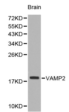
TargetVAMP2
Overview
- SupplierBioss
- Product NameVAMP2 Recombinant Antibody
- Delivery Days Customer16
- ApplicationsFlow Cytometry, ImmunoFluorescence, Western Blot, ImmunoCytoChemistry, ImmunoHistoChemistry, ImmunoHistoChemistry Paraffin
- Applications SupplierWB(1:300-5000), FCM(1:20-100), IHC-P(1:200-400), IF(ICC)(1:50-200)
- CertificationResearch Use Only
- ClonalityMonoclonal
- Concentration1 ug/ul
- ConjugateUnconjugated
- Gene ID6844
- Target nameVAMP2
- Target descriptionvesicle associated membrane protein 2
- Target synonymsNEDHAHM, SYB2, VAMP-2, vesicle-associated membrane protein 2, synaptobrevin 2
- HostRabbit
- IsotypeIgG
- Protein IDP63027
- Protein NameVesicle-associated membrane protein 2
- ReactivityHuman, Mouse, Rat
- Storage Instruction-20°C
- UNSPSC41116161
Datasheet
Related products
Product group Antibodies
Anti-VAMP2 AntibodyA29538
ApplicationsWestern Blot, ImmunoHistoChemistry
ReactivityHuman, Mouse, Rat
- SizePrice
Product group Antibodies
Anti-VAMP2 Antibody144-60082
ApplicationsWestern Blot, ImmunoHistoChemistry
ReactivityHuman, Mouse, Rat
TargetVAMP2
- SizePrice
Product group Antibodies

VAMP2 / VAMP-2 AntibodyLS-C771690
ApplicationsWestern Blot, ELISA, ImmunoHistoChemistry
ReactivityHuman, Mouse, Rat
TargetVAMP2
- SizePrice
Product group Antibodies
VAMP2 AntibodyCSB-PA025781ESR2HU
ApplicationsELISA, ImmunoHistoChemistry
ReactivityHuman
TargetVAMP2
- SizePrice

Product group Antibodies
ApplicationsFlow Cytometry, ImmunoFluorescence, ImmunoPrecipitation, Western Blot, ImmunoCytoChemistry
ReactivityHuman, Mouse, Rat
TargetVAMP2
- SizePrice
![VAMP2 antibody detects VAMP2 protein at synaptic vesicles by immunofluorescent analysis. Sample: DIV9 rat E18 primary cortical neurons were fixed in 4% paraformaldehyde at RT for 15 min. Green: VAMP2 protein stained by VAMP2 antibody (GTX121462) diluted at 1:500. Red: beta Tubulin 3/ Tuj1, stained by beta Tubulin 3/ Tuj1 antibody [GT11710] (GTX631836) diluted at 1:500. Blue: Fluoroshield with DAPI (GTX30920).](https://www.genetex.com/upload/website/prouct_img/normal/GTX121462/GTX121462_40513_20170503_IFA_R_w_23060519_797.webp)
Product group Antibodies
VAMP2 antibodyGTX121462
ApplicationsImmunoFluorescence, Western Blot, ImmunoCytoChemistry
ReactivityHuman, Mouse, Rat
TargetVAMP2
- SizePrice
Product group Antibodies
ApplicationsImmunoFluorescence, Western Blot, ELISA, ImmunoCytoChemistry
ReactivityHuman
TargetVAMP2
- SizePrice