

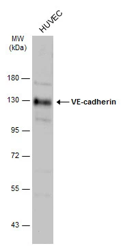
Whole cell extract (30 μg) was separated by 7.5% SDS-PAGE, and the membrane was blotted with VE-cadherin antibody (GTX132982) diluted at 1:2000. The HRP-conjugated anti-rabbit IgG antibody (GTX213110-01) was used to detect the primary antibody.
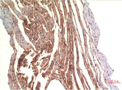
 diluted at 1:500. Antigen Retrieval: Citrate buffer, pH 6.0, 15 min")
Whole cell extract (30 μg) was separated by 7.5% SDS-PAGE, and the membrane was blotted with VE-cadherin antibody (GTX132982) diluted at 1:2000. The HRP-conjugated anti-rabbit IgG antibody (GTX213110-01) was used to detect the primary antibody.
VE-Cadherin antibody
GTX132982
ApplicationsWestern Blot, ImmunoHistoChemistry, ImmunoHistoChemistry Paraffin
Product group Antibodies
ReactivityHuman
TargetCDH5
Overview
- SupplierGeneTex
- Product NameVE-Cadherin antibody
- Delivery Days Customer9
- Application Supplier NoteWB: 1:500-1:3000. *Optimal dilutions/concentrations should be determined by the researcher.Not tested in other applications.
- ApplicationsWestern Blot, ImmunoHistoChemistry, ImmunoHistoChemistry Paraffin
- CertificationResearch Use Only
- ClonalityPolyclonal
- Concentration1.56 mg/ml
- ConjugateUnconjugated
- Gene ID1003
- Target nameCDH5
- Target descriptioncadherin 5
- Target synonyms7B4, CD144, cadherin-5, 7B4 antigen, VE-cadherin, cadherin 5, type 2 (vascular endothelium), cadherin 5, type 2, VE-cadherin (vascular epithelium), cd144 antigen, endothelial-specific cadherin, vascular endothelial cadherin
- HostRabbit
- IsotypeIgG
- Protein IDP33151
- Protein NameCadherin-5
- Scientific DescriptionThis gene is a classical cadherin from the cadherin superfamily and is located in a six-cadherin cluster in a region on the long arm of chromosome 16 that is involved in loss of heterozygosity events in breast and prostate cancer. The encoded protein is a calcium-dependent cell-cell adhesion glycoprotein comprised of five extracellular cadherin repeats, a transmembrane region and a highly conserved cytoplasmic tail. Functioning as a classic cadherin by imparting to cells the ability to adhere in a homophilic manner, the protein may play an important role in endothelial cell biology through control of the cohesion and organization of the intercellular junctions. An alternative splice variant has been described but its full length sequence has not been determined. [provided by RefSeq]
- ReactivityHuman
- Storage Instruction-20°C or -80°C,2°C to 8°C
- UNSPSC41116161
Datasheet
Related products

Product group Antibodies
Anti-CDH5 AntibodyA98230
ApplicationsWestern Blot, ELISA
ReactivityHuman, Mouse, Rat
- SizePrice
Product group Antibodies

ApplicationsWestern Blot, ELISA, ImmunoHistoChemistry
- SizePrice

Product group Antibodies
Anti-VE-Cadherin CDH5-Antibody Picoband(r)A02632-1-CARRIER-FREE
ApplicationsFlow Cytometry, ImmunoFluorescence, Western Blot, ELISA, ImmunoHistoChemistry
ReactivityHuman
TargetCDH5
- SizePrice
Product group Antibodies
Anti-CDH5 Antibody144-00734
ApplicationsWestern Blot
ReactivityHuman, Mouse, Rat
TargetCDH5
- SizePrice
Product group Antibodies
ApplicationsFlow Cytometry, Western Blot
ReactivityHuman, Mouse, Rat
TargetCDH5
- SizePrice
Product group Antibodies
CDH5 Monoclonal AntibodyCSB-MA249655
ApplicationsELISA, ImmunoHistoChemistry
ReactivityHuman, Mouse, Rat
TargetCDH5
- SizePrice
Product group Antibodies

Goat anti-VE-cadherinEB12667
ApplicationsFlow Cytometry, ImmunoFluorescence, ELISA
ReactivityBovine, Human, Porcine
TargetCDH5
- SizePrice
Product group Antibodies
Cdh5 Polyclonal AntibodyCAC08147
ApplicationsImmunoFluorescence, Western Blot, ELISA
ReactivityMouse
TargetCDH5
- SizePrice

Product group Antibodies
Anti-CDH5 AntibodyHPA075875
ApplicationsWestern Blot, ImmunoCytoChemistry
ReactivityHuman
TargetCDH5
- SizePrice