

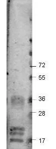
Western blot using GeneTex's Protein-A Purified anti-bovine VEGF antibody shows detection of recombinant bovine VEGF-A at 17-19.2 kDa. Approximately 2 μg of recombinant protein was loaded per lane onto a 4-20% gradient gel followed by transfer to PVDF membrane. The membrane was blocked using 3% BSA diluted 1:10. The primary antibody was used at a 1:333 dilution and was incubated with the blot for 2h at room temperature. The membrane was washed and reacted with a 1:10,000 dilution of IRDye800 Conjugated Affinity Purified Goat-anti-Rabbit IgG [H&L] MX10. Molecular weight estimation was made by comparison to prestained MW markers. Other detection systems will yield similar results.

Western blot using GeneTex's Protein-A Purified anti-bovine VEGF antibody shows detection of recombinant bovine VEGF-A at 17-19.2 kDa. Approximately 2 μg of recombinant protein was loaded per lane onto a 4-20% gradient gel followed by transfer to PVDF membrane. The membrane was blocked using 3% BSA diluted 1:10. The primary antibody was used at a 1:333 dilution and was incubated with the blot for 2h at room temperature. The membrane was washed and reacted with a 1:10,000 dilution of IRDye800 Conjugated Affinity Purified Goat-anti-Rabbit IgG [H&L] MX10. Molecular weight estimation was made by comparison to prestained MW markers. Other detection systems will yield similar results.
VEGFA antibody
GTX48811
ApplicationsWestern Blot, ELISA
Product group Antibodies
ReactivityBovine
TargetVEGFA
Overview
- SupplierGeneTex
- Product NameVEGFA antibody
- Delivery Days Customer9
- Application Supplier NoteWB: 1:333. *Optimal dilutions/concentrations should be determined by the researcher.Not tested in other applications.
- ApplicationsWestern Blot, ELISA
- CertificationResearch Use Only
- ClonalityPolyclonal
- Concentration1 mg/ml
- ConjugateUnconjugated
- Gene ID281572
- Target nameVEGFA
- Target descriptionvascular endothelial growth factor A
- Target synonymsVEGF, VEGF-A, VPF, eVEGF120, eVEGF164, vascular endothelial growth factor A, vascular endothelial growth factor 164, vascular permeability factor
- HostRabbit
- IsotypeIgG
- Protein IDP15691
- Protein NameVascular endothelial growth factor A
- Scientific DescriptionVEGF is a potent mitogen in embryonic and somatic angiogenesis with specificity for vascular endothelial cells. VEGF forms homodimers and exists in four different isoforms. Overall, the VEGF monomer resembles that of PDGF, but its N-terminal segment is helical rather than extended. VEGF shares homologies of about 21% and 24% respectively with the A and B chains of human platelet-derived growth factor (PDGF), and has complete conservation of the eight cysteine residues found in both mature PDGF chains. The cysteine knot motif is a common feature of this domain. The homology is not reflected in function, however, since the cell types responsive to VEGF are distinct from those responsive to homo- and heterodimers of the PDGF chains. This protein is a glycosylated mitogen that acts on endothelial cells and has various effects, including mediating increased vascular permeability, inducing angiogenesis, vasculogenesis and endothelial cell growth, promoting cell migration, and inhibiting apoptosis. VEGF-A also has been shown to have effects on a number of other cell types (e.g. stimulation of monocyte/macrophage migration, neurons, cancer cells, kidney epithelial cells ). VEGF-A is also a vasodilator; it increases microvascular permeability, and was originally referred to as vascular permeability factor. Alternatively spliced transcript variants, encoding either freely secreted or cell-associated isoforms, have been characterized.
- ReactivityBovine
- Storage Instruction-20°C or -80°C,2°C to 8°C
- UNSPSC41116161
References
- Leptin Promotes Angiogenesis via Pericyte STAT3 Pathway upon Intracerebral Hemorrhage.Read this paper
Datasheet
Related products
Product group Antibodies
ApplicationsImmunoPrecipitation, Western Blot, ImmunoCytoChemistry, ImmunoHistoChemistry
ReactivityBovine
TargetVEGFA
- SizePrice