

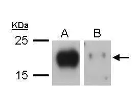
Sample (20 μg of whole cell lysate) A: TL1 whole cell lysate B: TL1 knock down VEGF-C whole cell lysate 12% SDS PAGE GTX113574 diluted at 1:1000 The HRP-conjugated anti-rabbit IgG antibody (GTX213110-01) was used to detect the primary antibody.
 antibody at 1:500 dilution.
Antigen Retrieval: Trilogy? (EDTA based, pH 8.0) buffer, 15min")
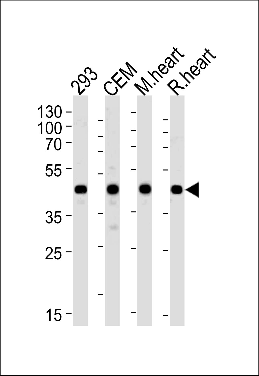
 was separated by 10% SDS-PAGE, and the membrane was blotted with VEGFC antibody (GTX113574) diluted at 1:1000. The HRP-conjugated anti-rabbit IgG antibody (GTX213110-01) was used to detect the primary antibody.")
 dilution: 1:1000 The HRP-conjugated anti-rabbit IgG antibody (GTX213110-01) was used to detect the primary antibody.")
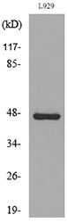
 were separated by 10% SDS-PAGE, and the membrane was blotted with VEGFC antibody (GTX113574) diluted at 1:500. The HRP-conjugated anti-rabbit IgG antibody (GTX213110-01) was used to detect the primary antibody.")
 diluted at 1:500. Antigen Retrieval: Citrate buffer, pH 6.0, 15 min")
 diluted at 1:500. Antigen Retrieval: Citrate buffer, pH 6.0, 15 min")
Sample (20 μg of whole cell lysate) A: TL1 whole cell lysate B: TL1 knock down VEGF-C whole cell lysate 12% SDS PAGE GTX113574 diluted at 1:1000 The HRP-conjugated anti-rabbit IgG antibody (GTX213110-01) was used to detect the primary antibody.
VEGFC antibody
GTX113574
ApplicationsWestern Blot, ImmunoHistoChemistry, ImmunoHistoChemistry Paraffin
Product group Antibodies
ReactivityBovine, Human, Mouse, Rat
TargetVEGFC
Overview
- SupplierGeneTex
- Product NameVEGFC antibody
- Delivery Days Customer9
- Application Supplier NoteWB: 1:500-1:3000. IHC-P: 1:100-1:1000. *Optimal dilutions/concentrations should be determined by the researcher.Not tested in other applications.
- ApplicationsWestern Blot, ImmunoHistoChemistry, ImmunoHistoChemistry Paraffin
- CertificationResearch Use Only
- ClonalityPolyclonal
- Concentration0.3 mg/ml
- ConjugateUnconjugated
- Gene ID7424
- Target nameVEGFC
- Target descriptionvascular endothelial growth factor C
- Target synonymsFlt4-L, LMPH1D, LMPHM4, VRP, vascular endothelial growth factor C, FLT4 ligand DHM, vascular endothelial growth factor-related protein
- HostRabbit
- IsotypeIgG
- Protein IDP49767
- Protein NameVascular endothelial growth factor C
- Scientific DescriptionThe protein encoded by this gene is a member of the platelet-derived growth factor/vascular endothelial growth factor (PDGF/VEGF) family, is active in angiogenesis and endothelial cell growth, and can also affect the permeability of blood vessels. This secreted protein undergoes a complex proteolytic maturation, generating multiple processed forms which bind and activate VEGFR-3 receptors. Only the fully processed form can bind and activate VEGFR-2 receptors. This protein is structurally and functionally similar to vascular endothelial growth factor D. [provided by RefSeq]
- ReactivityBovine, Human, Mouse, Rat
- Storage Instruction-20°C or -80°C,2°C to 8°C
- UNSPSC41116161
Datasheet
Related products
Product group Antibodies
Anti-VEGFC AntibodyA97111
ApplicationsWestern Blot, ELISA
ReactivityHuman, Mouse, Rat
- SizePrice
Product group Antibodies
Anti-VEGFC Antibody144-02556
ApplicationsWestern Blot, ImmunoHistoChemistry
ReactivityHuman, Mouse, Rat
TargetVEGFC
- SizePrice
Product group Antibodies

VEGFC AntibodyLS-C747629
ApplicationsWestern Blot, ImmunoHistoChemistry
ReactivityHuman, Mouse
TargetVEGFC
- SizePrice

Product group Antibodies
Anti-VEGFC AntibodyM00623
ApplicationsWestern Blot
ReactivityHuman, Mouse, Rat
TargetVEGFC
- SizePrice
Product group Antibodies
VEGF-C (7C4) Monoclonal AntibodyBSM-51054M
ApplicationsWestern Blot
ReactivityHuman, Mouse, Rat
TargetVEGFC
- SizePrice
Product group Antibodies
VEGFC Polyclonal AntibodyCAC14101
ApplicationsWestern Blot, ELISA, ImmunoHistoChemistry
TargetVEGFC
- SizePrice

Product group Antibodies
VEGFC AntibodyCSB-PA005364
ApplicationsWestern Blot, ELISA
ReactivityHuman, Mouse, Rat
TargetVEGFC
- SizePrice
![IHC-P analysis of formalin fixed human placenta tissue using GTX52459 VEGFC antibody [2E65].](https://www.genetex.com/upload/website/prouct_img/normal/GTX52459/GTX52459_20191119_IHC-P_w_23060900_778.webp)
Product group Antibodies
VEGFC antibody [2E65]GTX52459
ApplicationsWestern Blot, ImmunoHistoChemistry, ImmunoHistoChemistry Paraffin
ReactivityHuman
TargetVEGFC
- SizePrice
![VEGFC antibody [HL2289] detects VEGFC protein at cytoplasmic vesicles by immunofluorescent analysis. Sample: U87-MG cells were fixed in 4% paraformaldehyde at RT for 15 min. Green: VEGFC stained by VEGFC antibody [HL2289] (GTX638344) diluted at 1:500. Red: alpha Tubulin, a cytoskeleton marker, stained by alpha Tubulin antibody [GT114] (GTX628802) diluted at 1:1000. Blue: Fluoroshield with DAPI (GTX30920).](https://www.genetex.com/upload/website/prouct_img/normal/GTX638344/GTX638344_T-44984_20230421_ICC_IF_23050918_176.webp)
Product group Antibodies
VEGFC antibody [HL2289]GTX638344
ApplicationsImmunoFluorescence, Western Blot, ImmunoCytoChemistry
ReactivityHuman
TargetVEGFC
- SizePrice
Product group Antibodies
VEGFC antibodyGTX59592
ApplicationsWestern Blot, ImmunoHistoChemistry, ImmunoHistoChemistry Frozen, ImmunoHistoChemistry Paraffin
ReactivityHuman
TargetVEGFC
- SizePrice