

VGAT antibody [N1N2], N-term detects VGAT protein expression by immunofluorescent analysis. Sample: Cultured rat E18 primary cortical neuron, DIV 8. Cells were fixed in 4% paraformaldehyde at RT for 15 min. Green: VGAT protein stained by VGAT antibody [N1N2], N-term (GTX101908) diluted at 1:250. Red: beta Tubulin 3/ TUJ1, stained by beta Tubulin 3/ TUJ1 antibody [GT11710] (GTX631836) diluted at 1:250. Blue: Fluoroshield with DAPI (GTX30920).
![VGAT antibody [N1N2], N-term detects VGAT protein expression by immunohistochemical analysis. Sample: Frozen sectioned adult mouse retina. Green: VGAT protein stained by VGAT antibody [N1N2], N-term (GTX101908) diluted at 1:250. Red: beta Tubulin 3/ TUJ1, stained by beta Tubulin 3/ TUJ1 antibody [GT11710] (GTX631836) diluted at 1:250. Blue: Fluoroshield with DAPI (GTX30920).](https://www.genetex.com/upload/website/prouct_img/normal/GTX101908/GTX101908_40275_20170214_IHC-Fr_w_23060100_881.webp "VGAT antibody [N1N2], N-term detects VGAT protein expression by immunohistochemical analysis. Sample: Frozen sectioned adult mouse retina. Green: VGAT protein stained by VGAT antibody [N1N2], N-term (GTX101908) diluted at 1:250. Red: beta Tubulin 3/ TUJ1, stained by beta Tubulin 3/ TUJ1 antibody [GT11710] (GTX631836) diluted at 1:250. Blue: Fluoroshield with DAPI (GTX30920).")
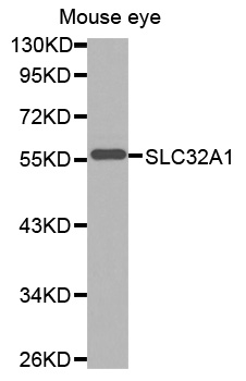
![VGAT antibody [N1N2], N-term detects VGAT protein at cytoplasm by immunohistochemical analysis. Sample: Paraffin-embedded mouse eye. Green: VGAT stained by VGAT antibody [N1N2], N-term (GTX101908) diluted at 1:500. Red: beta Tubulin 3/ Tuj1, a neural marker, stained by beta Tubulin 3/ Tuj1 antibody [GT11710] (GTX631836) diluted at 1:500. Blue: Fluoroshield with DAPI (GTX30920). Antigen Retrieval: Citrate buffer, pH 6.0, 15 min](https://www.genetex.com/upload/website/prouct_img/normal/GTX101908/GTX101908_44567_20220415_IHC-P_M_w_23060100_706.webp "VGAT antibody [N1N2], N-term detects VGAT protein at cytoplasm by immunohistochemical analysis. Sample: Paraffin-embedded mouse eye. Green: VGAT stained by VGAT antibody [N1N2], N-term (GTX101908) diluted at 1:500. Red: beta Tubulin 3/ Tuj1, a neural marker, stained by beta Tubulin 3/ Tuj1 antibody [GT11710] (GTX631836) diluted at 1:500. Blue: Fluoroshield with DAPI (GTX30920). Antigen Retrieval: Citrate buffer, pH 6.0, 15 min")
![VGAT antibody [N1N2], N-term detects VGAT protein expression by immunohistochemical analysis. Sample: Frozen-sectioned adult mouse cerebellum. Green: VGAT protein stained by VGAT antibody [N1N2], N-term (GTX101908) diluted at 1:250. Red: beta Tubulin 3/ TUJ1, stained by beta Tubulin 3/ TUJ1 antibody [GT11710] (GTX631836) diluted at 1:500. Blue: Fluoroshield with DAPI (GTX30920).](https://www.genetex.com/upload/website/prouct_img/normal/GTX101908/GTX101908_40275_20170531_IHC-Fr_M_w_23060100_295.webp "VGAT antibody [N1N2], N-term detects VGAT protein expression by immunohistochemical analysis. Sample: Frozen-sectioned adult mouse cerebellum. Green: VGAT protein stained by VGAT antibody [N1N2], N-term (GTX101908) diluted at 1:250. Red: beta Tubulin 3/ TUJ1, stained by beta Tubulin 3/ TUJ1 antibody [GT11710] (GTX631836) diluted at 1:500. Blue: Fluoroshield with DAPI (GTX30920).")
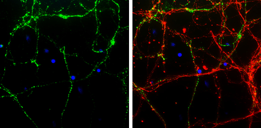
![VGAT antibody [N1N2], N-term detects VGAT protein by immunofluorescent analysis.
Sample: DIV9 rat cortical neuron and Glia cells were fixed in 4% paraformaldehyde at RT for 15 min.
Green: VGAT stained by VGAT antibody [N1N2], N-term (GTX101908) diluted at 1:250.
Red: Tau, an axon marker, stained by Tau antibody [GT287] (GTX634809) diluted at 1:500.](https://www.genetex.com/upload/website/prouct_img/normal/GTX101908/GTX101908_43971_20211112_ICC_IF_R_w_23060100_310.webp "VGAT antibody [N1N2], N-term detects VGAT protein by immunofluorescent analysis.
Sample: DIV9 rat cortical neuron and Glia cells were fixed in 4% paraformaldehyde at RT for 15 min.
Green: VGAT stained by VGAT antibody [N1N2], N-term (GTX101908) diluted at 1:250.
Red: Tau, an axon marker, stained by Tau antibody [GT287] (GTX634809) diluted at 1:500.")
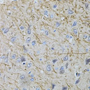
 antibody at 1:500 dilution.
Antigen Retrieval: Trilogy? (EDTA based, pH 8.0) buffer, 15min")
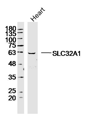
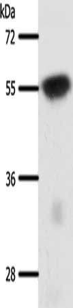
![Boiled and unboiled various tissue extracts (50 μg) were separated by 10% SDS-PAGE, and the membrane was blotted with VGAT antibody [N1N2], N-term (GTX101908) diluted at 1:3000. The HRP-conjugated anti-rabbit IgG antibody (GTX213110-01) was used to detect the primary antibody.](https://www.genetex.com/upload/website/prouct_img/normal/GTX101908/GTX101908_45133_20230818_WB_M_brain_25021320_499.webp "Boiled and unboiled various tissue extracts (50 μg) were separated by 10% SDS-PAGE, and the membrane was blotted with VGAT antibody [N1N2], N-term (GTX101908) diluted at 1:3000. The HRP-conjugated anti-rabbit IgG antibody (GTX213110-01) was used to detect the primary antibody.")
![Non-transfected (–) and transfected (+) unboiled 293T whole cell extracts (30 μg) were separated by 10% SDS-PAGE, and the membrane was blotted with VGAT antibody [N1N2], N-term (GTX101908) diluted at 1:10000. The HRP-conjugated anti-rabbit IgG antibody (GTX213110-01) was used to detect the primary antibody.](https://www.genetex.com/upload/website/prouct_img/normal/GTX101908/GTX101908_45133_20230818_WB_B_25021320_686.webp "Non-transfected (–) and transfected (+) unboiled 293T whole cell extracts (30 μg) were separated by 10% SDS-PAGE, and the membrane was blotted with VGAT antibody [N1N2], N-term (GTX101908) diluted at 1:10000. The HRP-conjugated anti-rabbit IgG antibody (GTX213110-01) was used to detect the primary antibody.")
VGAT antibody [N1N2], N-term detects VGAT protein expression by immunofluorescent analysis. Sample: Cultured rat E18 primary cortical neuron, DIV 8. Cells were fixed in 4% paraformaldehyde at RT for 15 min. Green: VGAT protein stained by VGAT antibody [N1N2], N-term (GTX101908) diluted at 1:250. Red: beta Tubulin 3/ TUJ1, stained by beta Tubulin 3/ TUJ1 antibody [GT11710] (GTX631836) diluted at 1:250. Blue: Fluoroshield with DAPI (GTX30920).
VGAT antibody [N1N2], N-term
GTX101908
ApplicationsImmunoFluorescence, Western Blot, ImmunoCytoChemistry, ImmunoHistoChemistry, ImmunoHistoChemistry Frozen, ImmunoHistoChemistry Paraffin
Product group Antibodies
ReactivityHuman, Mouse, Rat
TargetSLC32A1
Overview
- SupplierGeneTex
- Product NameVGAT antibody [N1N2], N-term
- Delivery Days Customer9
- Application Supplier NoteWB: 1:1000-1:10000. ICC/IF: 1:100-1:1000. IHC-P: 1:100-1:1000. IHC-Fr: 1:100-1:1000. *Optimal dilutions/concentrations should be determined by the researcher.Not tested in other applications.
- ApplicationsImmunoFluorescence, Western Blot, ImmunoCytoChemistry, ImmunoHistoChemistry, ImmunoHistoChemistry Frozen, ImmunoHistoChemistry Paraffin
- CertificationResearch Use Only
- ClonalityPolyclonal
- Concentration0.52 mg/ml
- ConjugateUnconjugated
- Gene ID140679
- Target nameSLC32A1
- Target descriptionsolute carrier family 32 member 1
- Target synonymsDEE114, GEFSP12, VGAT, VIAAT, VIAAT GEFSP12, vesicular inhibitory amino acid transporter, GABA and glycine transporter, hVIAAT, solute carrier family 32 (GABA vesicular transporter), member 1, vesicular GABA transporter
- HostRabbit
- IsotypeIgG
- Protein IDQ9H598
- Protein NameVesicular inhibitory amino acid transporter
- Scientific DescriptionThe protein encoded by this gene is an integral membrane protein involved in gamma-aminobutyric acid (GABA) and glycine uptake into synaptic vesicles. The encoded protein is a member of amino acid/polyamine transporter family II. [provided by RefSeq]
- ReactivityHuman, Mouse, Rat
- Storage Instruction-20°C or -80°C,2°C to 8°C
- UNSPSC41116161
Datasheet
Related products
Product group Antibodies
Anti-SLC32A1 AntibodyA30706
ApplicationsWestern Blot, ImmunoHistoChemistry
ReactivityHuman, Mouse, Rat
- SizePrice
Product group Antibodies
Anti-Mouse/Rat SLC32A1 Antibody144-03129
ApplicationsWestern Blot, ImmunoHistoChemistry
ReactivityHuman, Mouse, Rat
TargetSLC32A1
- SizePrice
Product group Antibodies
SLC32A1 Polyclonal AntibodyBS-19821R
ApplicationsImmunoFluorescence, Western Blot, ELISA, ImmunoCytoChemistry, ImmunoHistoChemistry, ImmunoHistoChemistry Frozen, ImmunoHistoChemistry Paraffin
ReactivityBovine, Human, Mouse, Porcine, Rabbit, Rat
TargetSLC32A1
- SizePrice
Product group Antibodies
Slc32A1 Polyclonal AntibodyCAC11154
ApplicationsImmunoFluorescence, Western Blot, ELISA, ImmunoHistoChemistry
ReactivityMouse
TargetSLC32A1
- SizePrice
Product group Antibodies
SLC32A1 AntibodyCSB-PA034916
ApplicationsWestern Blot, ELISA
ReactivityHuman, Mouse, Rat
TargetSLC32A1
- SizePrice
Product group Antibodies

SLC32A1 / VGAT AntibodyLS-C402787
ApplicationsWestern Blot, ELISA
ReactivityHuman, Mouse, Rat
TargetSLC32A1
- SizePrice

Product group Antibodies
Anti-VGAT AntibodyHPA058859
ApplicationsImmunoHistoChemistry
ReactivityHuman
TargetSLC32A1
- SizePrice
![VGAT antibody [HL1615] detects VGAT protein at cytoplasm by immunohistochemical analysis. Sample: Paraffin-embedded mouse cerebellum. VGAT stained by VGAT antibody [HL1615] (GTX637106) diluted at 1:100. Antigen Retrieval: Citrate buffer, pH 6.0, 15 min](https://www.genetex.com/upload/website/prouct_img/normal/GTX637106/GTX637106_T-44732_20220805_IHC-P_M_22080923_282.webp)
Product group Antibodies
VGAT antibody [HL1615]GTX637106
ApplicationsWestern Blot, ImmunoHistoChemistry, ImmunoHistoChemistry Frozen, ImmunoHistoChemistry Paraffin
ReactivityHuman, Mouse
TargetSLC32A1
- SizePrice
![VGAT antibody [HL1616] detects VGAT protein at cytoplasm by immunohistochemical analysis. Sample: Paraffin-embedded mouse cerebellum. VGAT stained by VGAT antibody [HL1616] (GTX637107) diluted at 1:100. Antigen Retrieval: Citrate buffer, pH 6.0, 15 min](https://www.genetex.com/upload/website/prouct_img/normal/GTX637107/GTX637107_T-44732_20220805_IHC-P_M_22080923_105.webp)
Product group Antibodies
VGAT antibody [HL1616]GTX637107
ApplicationsImmunoFluorescence, Western Blot, ImmunoCytoChemistry, ImmunoHistoChemistry, ImmunoHistoChemistry Frozen, ImmunoHistoChemistry Paraffin
ReactivityHuman, Mouse, Rat
TargetSLC32A1
- SizePrice
Product group Antibodies
VGAT antibodyGTX32964
ApplicationsImmunoFluorescence, Western Blot, ImmunoCytoChemistry, ImmunoHistoChemistry, ImmunoHistoChemistry Paraffin
ReactivityHuman, Mouse, Rat
TargetSLC32A1
- SizePrice