

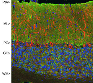
Confocal image of adult rat cerebellar cortex stained with VSNL1 (green) and chicken polyclonal antibody to MAP2 (red) and DNA (blue). VSNL1 antibody reveals synapses in the molecular layer (ML) strongly. Synaptic regions are also seen in the granule cell layer (GC). The perikarya of Purkinje cells (PC) are revealed with the MAP2 antibody (4). Little staining is seen in the white matter (WM). Protocol on Data-sheet.
Confocal image of adult rat cerebellar cortex stained with VSNL1 (green) and chicken polyclonal antibody to MAP2 (red) and DNA (blue). VSNL1 antibody reveals synapses in the molecular layer (ML) strongly. Synaptic regions are also seen in the granule cell layer (GC). The perikarya of Purkinje cells (PC) are revealed with the MAP2 antibody (4). Little staining is seen in the white matter (WM). Protocol on Data-sheet.
VILIP1 (VSNL1) Mouse Monoclonal Antibody
MO22132-100
ApplicationsImmunoFluorescence, Western Blot
Product group Antibodies
ReactivityBovine, Human, Mouse, Rat
TargetVSNL1
Overview
- SupplierOriGene
- Product NameVILIP1 (VSNL1) Mouse Monoclonal Antibody
- Delivery Days Customer14
- ApplicationsImmunoFluorescence, Western Blot
- CertificationResearch Use Only
- ClonalityMonoclonal
- Gene ID7447
- Target nameVSNL1
- Target descriptionvisinin like 1
- Target synonymsHLP3, HPCAL3, HUVISL1, VILIP, VILIP-1, visinin-like protein 1, VLP-1, hippocalcin-like protein 3
- HostMouse
- IsotypeIgG1
- Protein IDP62760
- Protein NameVisinin-like protein 1
- Scientific DescriptionVILIP1 (VSNL1) mouse monoclonal antibody
- ReactivityBovine, Human, Mouse, Rat
- UNSPSC12352203
Related products
Product group Antibodies

ApplicationsWestern Blot
- SizePrice
Product group Antibodies
Anti-VSNL1 Antibody144-02797
ApplicationsImmunoFluorescence, Western Blot
ReactivityHuman, Mouse, Rat
TargetVSNL1
- SizePrice
Product group Antibodies
ApplicationsImmunoPrecipitation, Western Blot, ImmunoCytoChemistry, ImmunoHistoChemistry
ReactivityMouse, Porcine, Rat
TargetVSNL1
- SizePrice
Product group Antibodies
VILIP1 Monoclonal AntibodyBSM-51682M
ApplicationsWestern Blot
ReactivityBovine, Chicken, Human, Mouse, Rat
TargetVSNL1
- SizePrice
Product group Antibodies
Anti-VSNL1 AntibodyA31803
ApplicationsImmunoFluorescence, Western Blot, ImmunoHistoChemistry
ReactivityHuman, Mouse, Rat
- SizePrice
Product group Antibodies

VILIP / VSNL1 AntibodyLS-C832546
ApplicationsWestern Blot, ELISA, ImmunoHistoChemistry
ReactivityHuman, Mouse, Rat
TargetVSNL1
- SizePrice
Product group Antibodies
VSNL1 AntibodyCSB-PA025933DSR2HU
ApplicationsELISA, ImmunoHistoChemistry
ReactivityHuman
TargetVSNL1
- SizePrice
Product group Antibodies
References
Visinin-like 1 antibodyGTX115039
ApplicationsWestern Blot, ImmunoHistoChemistry, ImmunoHistoChemistry Frozen, ImmunoHistoChemistry Paraffin
ReactivityHuman, Mouse, Rabbit, Rat
TargetVSNL1
- SizePrice