

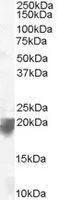
WB analysis of human spleen lysate using GTX10160 VPS29 antibody, C-term. Dilution : 0.1μg/ml Loading : 35μg protein in RIPA buffer
WB analysis of human spleen lysate using GTX10160 VPS29 antibody, C-term. Dilution : 0.1μg/ml Loading : 35μg protein in RIPA buffer
VPS29 antibody, C-term
GTX10160
ApplicationsWestern Blot
Product group Antibodies
ReactivityHuman
TargetVPS29
Overview
- SupplierGeneTex
- Product NameVPS29 antibody, C-term
- Delivery Days Customer7
- Application Supplier NoteWB: 0.1-0.3microg/ml. *Optimal dilutions/concentrations should be determined by the researcher.Not tested in other applications.
- ApplicationsWestern Blot
- CertificationResearch Use Only
- ClonalityPolyclonal
- Concentration0.50 mg/ml
- ConjugateUnconjugated
- Gene ID51699
- Target nameVPS29
- Target descriptionVPS29 retromer complex component
- Target synonymsDC15, DC7, PEP11, vacuolar protein sorting-associated protein 29, PEP11 homolog, epididymis secretory sperm binding protein, hVPS29, retromer protein, vacuolar protein sorting 29 homolog, vacuolar sorting protein VPS29/PEP11, vesicle protein sorting 29, x 007 protein
- HostGoat
- IsotypeIgG
- Protein IDQ9UBQ0
- Protein NameVacuolar protein sorting-associated protein 29
- Scientific DescriptionThis gene belongs to a group of vacuolar protein sorting (VPS) genes that, when functionally impaired, disrupt the efficient delivery of vacuolar hydrolases. The protein encoded by this gene is a component of a large multimeric complex, termed the retromer complex, which is involved in retrograde transport of proteins from endosomes to the trans-Golgi network. This VPS protein may be involved in the formation of the inner shell of the retromer coat for retrograde vesicles leaving the prevacuolar compartment. Alternative splice variants encoding different isoforms and representing non-protein coding transcripts have been found for this gene. [provided by RefSeq, Aug 2013]
- ReactivityHuman
- Storage Instruction-20°C or -80°C,2°C to 8°C
- UNSPSC41116161
References
- Regulation of retromer recruitment to endosomes by sequential action of Rab5 and Rab7. Rojas R et al., 2008 Nov 3, J Cell BiolRead this paper
Datasheet
Related products


Product group Antibodies
Anti-VPS29 AntibodyA88708
ApplicationsImmunoFluorescence, Western Blot, ImmunoCytoChemistry
ReactivityHuman, Mouse, Rat
- SizePrice
Product group Antibodies

VPS29 AntibodyLS-C748161
ApplicationsImmunoFluorescence, Western Blot
ReactivityHuman
TargetVPS29
- SizePrice
Product group Antibodies

Goat anti-VPS29 (C Terminus)EB06257
ApplicationsWestern Blot, ELISA
ReactivityBovine, Canine, Human, Mouse, Rat
TargetVPS29
- SizePrice

Product group Antibodies
Anti-VPS29 AntibodyA04097
ApplicationsImmunoFluorescence, Western Blot, ImmunoCytoChemistry
ReactivityHuman, Mouse, Rat
TargetVPS29
- SizePrice
Product group Antibodies
VPS29 AntibodyCSB-PA890661LA01HU
ApplicationsImmunoFluorescence, Western Blot, ELISA, ImmunoHistoChemistry
ReactivityHuman
TargetVPS29
- SizePrice
Product group Antibodies
Vps29 Polyclonal AntibodyCAC08087
ApplicationsWestern Blot, ELISA
TargetVPS29
- SizePrice
Product group Antibodies
VPS29 antibodyGTX65880
ApplicationsWestern Blot
ReactivityHuman
TargetVPS29
- SizePrice
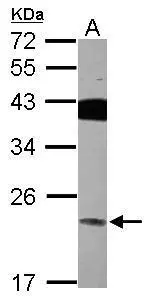
![Whole cell extract (30 μg) was separated by 12% SDS-PAGE, and the membrane was blotted with VPS29 antibody [C1C3] (GTX104768) diluted at 1:1500. The HRP-conjugated anti-rabbit IgG antibody (GTX213110-01) was used to detect the primary antibody, and the signal was developed with Trident ECL plus-Enhanced.](https://www.genetex.com/upload/website/prouct_img/normal/GTX104768/GTX104768_39666_20201211_WB_w_23060120_667.webp)
Product group Antibodies
VPS29 antibody [C1C3]GTX104768
ApplicationsWestern Blot
ReactivityHuman
TargetVPS29
- SizePrice
Product group Antibodies
VPS29 antibodyGTX116189
ApplicationsWestern Blot, ImmunoHistoChemistry, ImmunoHistoChemistry Paraffin
ReactivityHuman
TargetVPS29
- SizePrice