

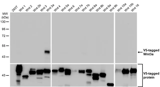
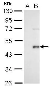
Non-transfected (–) and transfected (+) 293T whole cell extracts were separated by 10% SDS-PAGE, and the membrane was blotted with Wnt3a antibody [HL1911] (GTX637660) diluted at 1:5000. The HRP-conjugated anti-rabbit IgG antibody (GTX213110-01) was used to detect the primary antibody.
![Mouse tissue extract (50 μg) was separated by 10% SDS-PAGE, and the membrane was blotted with Wnt3a antibody [HL1911] (GTX637660) diluted at 1:1000. The HRP-conjugated anti-rabbit IgG antibody (GTX213110-01) was used to detect the primary antibody.](https://www.genetex.com/upload/website/prouct_img/normal/GTX637660/GTX637660_T-44837_20230804_WB_M_skin_23080901_299.webp "Mouse tissue extract (50 μg) was separated by 10% SDS-PAGE, and the membrane was blotted with Wnt3a antibody [HL1911] (GTX637660) diluted at 1:1000. The HRP-conjugated anti-rabbit IgG antibody (GTX213110-01) was used to detect the primary antibody.")
![Non-transfected (–) and transfected (+) 293T whole cell extracts (30 μg) were separated by 10% SDS-PAGE, and the membrane was blotted with Wnt3a antibody [HL1911] (GTX637660) diluted at 1:5000. The HRP-conjugated anti-rabbit IgG antibody (GTX213110-01) was used to detect the primary antibody, and the signal was developed with Trident ECL plus-Enhanced.](https://www.genetex.com/upload/website/prouct_img/normal/GTX637660/GTX637660_45138_20230825_WB_B_23083020_856.webp "Non-transfected (–) and transfected (+) 293T whole cell extracts (30 μg) were separated by 10% SDS-PAGE, and the membrane was blotted with Wnt3a antibody [HL1911] (GTX637660) diluted at 1:5000. The HRP-conjugated anti-rabbit IgG antibody (GTX213110-01) was used to detect the primary antibody, and the signal was developed with Trident ECL plus-Enhanced.")
![Wnt3a antibody [HL1911] detects secreted Wnt3a protein by immunohistochemical analysis. Sample: Paraffin-embedded human prostate cancer. Wnt3a stained by Wnt3a antibody [HL1911] (GTX637660) diluted at 1:100. Antigen Retrieval: Citrate buffer, pH 6.0, 15 min](https://www.genetex.com/upload/website/prouct_img/normal/GTX637660/GTX637660_45138_20230928_IHC-P_23103019_227.webp "Wnt3a antibody [HL1911] detects secreted Wnt3a protein by immunohistochemical analysis. Sample: Paraffin-embedded human prostate cancer. Wnt3a stained by Wnt3a antibody [HL1911] (GTX637660) diluted at 1:100. Antigen Retrieval: Citrate buffer, pH 6.0, 15 min")
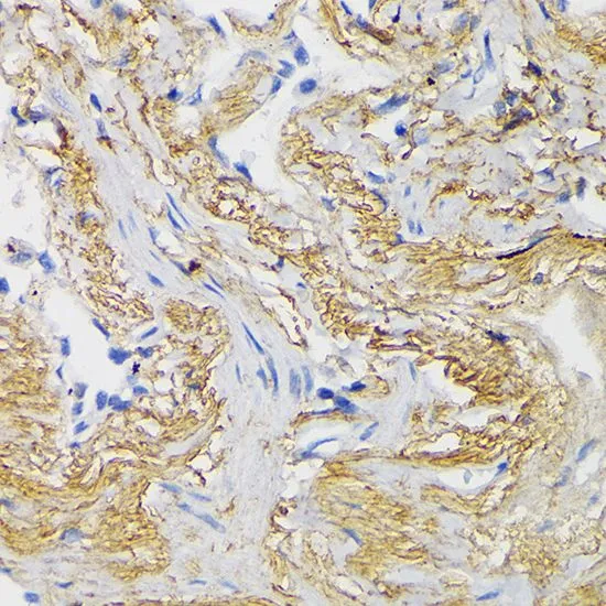
![Wnt3a antibody [HL1911] detects Wnt3a protein by immunohistochemical analysis. Sample: Paraffin-embedded rat tissues. Wnt3a stained by Wnt3a antibody [HL1911] (GTX637660) diluted at 1:200. Antigen Retrieval: Citrate buffer, pH 6.0, 15 min](https://www.genetex.com/upload/website/prouct_img/normal/GTX637660/GTX637660_45138_20231106_IHC-P_multiple_R_23111422_465.webp "Wnt3a antibody [HL1911] detects Wnt3a protein by immunohistochemical analysis. Sample: Paraffin-embedded rat tissues. Wnt3a stained by Wnt3a antibody [HL1911] (GTX637660) diluted at 1:200. Antigen Retrieval: Citrate buffer, pH 6.0, 15 min")
![Wnt3a antibody [HL1911] detects Wnt3a protein by immunofluorescent analysis. Sample: Mock and transfected 293T cells were fixed in ice-cold MeOH for 5 min. Green: Wnt3a stained by Wnt3a antibody [HL1911] (GTX637660) diluted at 1:500. Blue: Fluoroshield with DAPI (GTX30920). Scale bar= 10μm.](https://www.genetex.com/upload/website/prouct_img/normal/GTX637660/GTX637660_45138_20231110_ICC_IF_B_2_23111422_688.webp "Wnt3a antibody [HL1911] detects Wnt3a protein by immunofluorescent analysis. Sample: Mock and transfected 293T cells were fixed in ice-cold MeOH for 5 min. Green: Wnt3a stained by Wnt3a antibody [HL1911] (GTX637660) diluted at 1:500. Blue: Fluoroshield with DAPI (GTX30920). Scale bar= 10μm.")
Non-transfected (–) and transfected (+) 293T whole cell extracts were separated by 10% SDS-PAGE, and the membrane was blotted with Wnt3a antibody [HL1911] (GTX637660) diluted at 1:5000. The HRP-conjugated anti-rabbit IgG antibody (GTX213110-01) was used to detect the primary antibody.
Wnt3a antibody [HL1911]
GTX637660
ApplicationsImmunoFluorescence, Western Blot, ImmunoCytoChemistry, ImmunoHistoChemistry, ImmunoHistoChemistry Paraffin
Product group Antibodies
ReactivityHuman, Mouse, Rat
TargetWNT3A
Overview
- SupplierGeneTex
- Product NameWnt3a antibody [HL1911]
- Delivery Days Customer9
- Application Supplier NoteWB: 1:1000-1:10000. *Optimal dilutions/concentrations should be determined by the researcher.Not tested in other applications.
- ApplicationsImmunoFluorescence, Western Blot, ImmunoCytoChemistry, ImmunoHistoChemistry, ImmunoHistoChemistry Paraffin
- CertificationResearch Use Only
- ClonalityMonoclonal
- Clone IDHL1911
- Concentration1 mg/ml
- ConjugateUnconjugated
- Gene ID89780
- Target nameWNT3A
- Target descriptionWnt family member 3A
- Target synonymsprotein Wnt-3a, wingless-type MMTV integration site family, member 3A
- HostRabbit
- IsotypeIgG
- Protein IDP56704
- Protein NameProtein Wnt-3a
- Scientific DescriptionThe WNT gene family consists of structurally related genes which encode secreted signaling proteins. These proteins have been implicated in oncogenesis and in several developmental processes, including regulation of cell fate and patterning during embryogenesis. This gene is a member of the WNT gene family. It encodes a protein which shows 96% amino acid identity to mouse Wnt3A protein, and 84% to human WNT3 protein, another WNT gene product. This gene is clustered with WNT14 gene, another family member, in chromosome 1q42 region. [provided by RefSeq, Jul 2008]
- ReactivityHuman, Mouse, Rat
- Storage Instruction-20°C or -80°C,2°C to 8°C
- UNSPSC41116161
Datasheet
Related products
Product group Antibodies
Anti-Wnt3a AntibodyA12665
ApplicationsWestern Blot
ReactivityHuman, Mouse, Rat
- SizePrice
Product group Antibodies
Anti-WNT3A Antibody144-00642
ApplicationsWestern Blot
ReactivityHuman, Mouse
TargetWNT3A
- SizePrice
Product group Antibodies
WNT3A Antibody, HRP conjugatedCSB-PA15549B0RB
ApplicationsELISA
ReactivityHuman
TargetWNT3A
- SizePrice
Product group Antibodies
ApplicationsImmunoPrecipitation, Western Blot, ImmunoCytoChemistry, ImmunoHistoChemistry
ReactivityBovine, Mouse, Rat
TargetWNT3A
- SizePrice
Product group Antibodies
References
Wnt3a Polyclonal AntibodyBS-1700R
ApplicationsImmunoFluorescence, Western Blot, ELISA, ImmunoCytoChemistry, ImmunoHistoChemistry, ImmunoHistoChemistry Frozen, ImmunoHistoChemistry Paraffin
ReactivityBovine, Canine, Human, Mouse, Porcine, Rabbit, Rat, Sheep
TargetWNT3A
- SizePrice
Product group Antibodies

WNT3A AntibodyLS-C401528
ApplicationsWestern Blot, ELISA, ImmunoHistoChemistry
ReactivityHuman, Mouse
TargetWNT3A
- SizePrice
Product group Antibodies
Wnt3a antibody [N1C3]GTX109037
ApplicationsWestern Blot
ReactivityHuman
TargetWNT3A
- SizePrice
Product group Antibodies
Wnt3a antibodyGTX128101
ApplicationsWestern Blot, ImmunoHistoChemistry, ImmunoHistoChemistry Paraffin
ReactivityHuman, Rat
TargetWNT3A
- SizePrice
![WB analysis of various sample lysates using GTX50033 Wnt3a antibody [3A6]. Lane 1 : 293T whole cell lysate Lane 2 : Wnt3a transfected 293T cell lysate Dilution : 1:1000 Loading : 40μg per lane](https://www.genetex.com/upload/website/prouct_img/normal/GTX50033/GTX50033_WB_1_w_23060823_177.webp)
Product group Antibodies
Wnt3a antibody [3A6]GTX50033
ApplicationsWestern Blot, ELISA, ImmunoHistoChemistry, ImmunoHistoChemistry Paraffin
ReactivityHuman
TargetWNT3A
- SizePrice
Product group Antibodies
Wnt3a antibodyGTX64367
ApplicationsWestern Blot, ImmunoHistoChemistry, ImmunoHistoChemistry Paraffin
ReactivityHuman, Mouse, Rat
TargetWNT3A
- SizePrice