

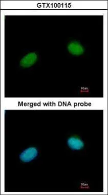
Immunofluorescence analysis of paraformaldehyde-fixed HeLa, using XRCC3(GTX100115) antibody at 1:200 dilution.
![Non-transfected (–) and transfected (+) 293T whole cell extracts (30 μg) were separated by 10% SDS-PAGE, and the membrane was blotted with XRCC3 antibody [C1C3] (GTX100115) diluted at 1:5000. The HRP-conjugated anti-rabbit IgG antibody (GTX213110-01) was used to detect the primary antibody.](https://www.genetex.com/upload/website/prouct_img/normal/GTX100115/GTX100115_39869_20230901_WB_B_23090619_210.webp "Non-transfected (–) and transfected (+) 293T whole cell extracts (30 μg) were separated by 10% SDS-PAGE, and the membrane was blotted with XRCC3 antibody [C1C3] (GTX100115) diluted at 1:5000. The HRP-conjugated anti-rabbit IgG antibody (GTX213110-01) was used to detect the primary antibody.")
Immunofluorescence analysis of paraformaldehyde-fixed HeLa, using XRCC3(GTX100115) antibody at 1:200 dilution.
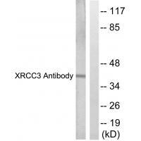
XRCC3 antibody [C1C3]
GTX100115
ApplicationsImmunoFluorescence, Western Blot, ImmunoCytoChemistry
Product group Antibodies
ReactivityHuman
TargetXRCC3
Overview
- SupplierGeneTex
- Product NameXRCC3 antibody [C1C3]
- Delivery Days Customer9
- Application Supplier NoteWB: 1:500-1:3000. ICC/IF: 1:100-1:1000. *Optimal dilutions/concentrations should be determined by the researcher.Not tested in other applications.
- ApplicationsImmunoFluorescence, Western Blot, ImmunoCytoChemistry
- CertificationResearch Use Only
- ClonalityPolyclonal
- Concentration1 mg/ml
- ConjugateUnconjugated
- Gene ID7517
- Target nameXRCC3
- Target descriptionX-ray repair cross complementing 3
- Target synonymsCMM6, DNA repair protein XRCC3, X-ray repair complementing defective repair in Chinese hamster cells 3, X-ray repair cross-complementing protein 3
- HostRabbit
- IsotypeIgG
- Protein IDO43542
- Protein NameDNA repair protein XRCC3
- Scientific DescriptionThis gene encodes a member of the RecA/Rad51-related protein family that participates in homologous recombination to maintain chromosome stability and repair DNA damage. This gene functionally complements Chinese hamster irs1SF, a repair-deficient mutant that exhibits hypersensitivity to a number of different DNA-damaging agents and is chromosomally unstable. A rare microsatellite polymorphism in this gene is associated with cancer in patients of varying radiosensitivity. Alternatively spliced transcript variants encoding the same protein have been identified. [provided by RefSeq]
- ReactivityHuman
- Storage Instruction-20°C or -80°C,2°C to 8°C
- UNSPSC41116161
Datasheet
Related products
Product group Antibodies
Anti-XRCC3 AntibodyA37710
ApplicationsWestern Blot, ImmunoHistoChemistry
ReactivityHuman
- SizePrice
Product group Antibodies
Anti-XRCC3 Antibody144-02134
ApplicationsImmunoFluorescence, Western Blot, ImmunoHistoChemistry
ReactivityHuman, Mouse, Rat
TargetXRCC3
- SizePrice
Product group Antibodies

Anti-XRCC3 [10F1/6]Ab01310-1.1
ApplicationsImmunoFluorescence, Western Blot, ImmunoCytoChemistry
ReactivityHuman
TargetXRCC3
- SizePrice

Product group Antibodies
XRCC3 Recombinant AntibodyBSM-62386R
ApplicationsFlow Cytometry, Western Blot
ReactivityHuman, Mouse, Rat
TargetXRCC3
- SizePrice
Product group Antibodies
XRCC3 AntibodyCSB-PA004531
ApplicationsWestern Blot, ELISA, ImmunoHistoChemistry
ReactivityHuman
TargetXRCC3
- SizePrice

Product group Antibodies
ApplicationsFlow Cytometry, Western Blot
ReactivityHuman, Mouse, Rat
TargetXRCC3
- SizePrice
Product group Antibodies
XRCC3 antibodyGTX26494
ApplicationsImmunoPrecipitation, Western Blot
ReactivityDrosophila, Human
TargetXRCC3
- SizePrice
![Non-transfected (–) and transfected (+) 293T whole cell extracts (30 μg) were separated by 10% SDS-PAGE, and the membrane was blotted with XRCC3 antibody [N1N3] (GTX100093) diluted at 1:5000. The HRP-conjugated anti-rabbit IgG antibody (GTX213110-01) was used to detect the primary antibody.](https://www.genetex.com/upload/website/prouct_img/normal/GTX100093/GTX100093_39408_20230901_WB_B_23090619_800.webp)
Product group Antibodies
XRCC3 antibody [N1N3]GTX100093
ApplicationsWestern Blot
ReactivityHuman
TargetXRCC3
- SizePrice
Product group Antibodies

XRCC3 AntibodyLS-C331928
ApplicationsImmunoFluorescence, Western Blot, ImmunoHistoChemistry
ReactivityHuman, Mouse, Rat
TargetXRCC3
- SizePrice