

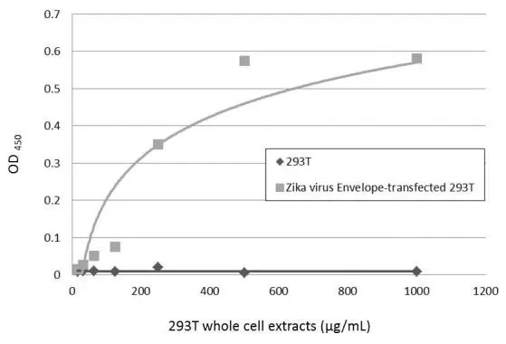
Sandwich ELISA detection of non-transfected and transfected 293T whole cell extracts using GTX634157 as capture antibody at concentration of 5 μg/mL and GTX133325 as detection antibody at concentration of 1 μg/mL. Rabbit IgG antibody (HRP) (GTX213110-01) was diluted at 1:10000 and used to detect the primary antibody.
 (GTX213110-01) was diluted at 1:10000 and used to detect the primary antibody.")
![Untreated (–) and treated (+) C6/36 cell extracts (15 μg) were separated by gradient gel, and the membrane was blotted with Zika virus Envelope protein antibody [GT871] (GTX634157) diluted at 1:2000.](https://www.genetex.com/upload/website/prouct_img/normal/GTX634157/GTX634157_42786_20170302_WB_Zika_w_23061202_746.webp "Untreated (–) and treated (+) C6/36 cell extracts (15 μg) were separated by gradient gel, and the membrane was blotted with Zika virus Envelope protein antibody [GT871] (GTX634157) diluted at 1:2000.")
![Immunofluorescent analysis of non-infected and infected vero cells using Zika virus Envelope protein antibody [GT871] (GTX634157). Green: Zika virus Envelope protein antibody [GT871] (GTX634157) diluted at 1:500.](https://www.genetex.com/upload/website/prouct_img/normal/GTX634157/GTX634157_42786_20170306_IFA_Zika_w_23061202_445.webp "Immunofluorescent analysis of non-infected and infected vero cells using Zika virus Envelope protein antibody [GT871] (GTX634157). Green: Zika virus Envelope protein antibody [GT871] (GTX634157) diluted at 1:500.")
 (GTX213111-01) was diluted at 1:10000 and used to detect the primary antibody.")
![Immunofluorescent analysis of arboviruses infected cells using Zika virus Envelope protein antibody [GT871] (GTX634157). Sample: Mock and zika virus-infected cells. Green: Zika virus Envelope protein antibody [GT871] (GTX634157) diluted at 1:100.](https://www.genetex.com/upload/website/prouct_img/normal/GTX634157/GTX634157_42786_20210903_ICC_IF_Zikavirus_w_23061202_774.webp "Immunofluorescent analysis of arboviruses infected cells using Zika virus Envelope protein antibody [GT871] (GTX634157). Sample: Mock and zika virus-infected cells. Green: Zika virus Envelope protein antibody [GT871] (GTX634157) diluted at 1:100.")
![Non-infected (–) and infected (+) Vero cell extracts (15 μg, unboiled) were separated by gradient gel, and the membrane was blotted with Zika virus Envelope protein antibody [GT871] (GTX634157) culture supernatant diluted at 1:50.](https://www.genetex.com/upload/website/prouct_img/normal/GTX634157/GTX634157_42786_20200826_WB_Zika_w_23061202_320.webp "Non-infected (–) and infected (+) Vero cell extracts (15 μg, unboiled) were separated by gradient gel, and the membrane was blotted with Zika virus Envelope protein antibody [GT871] (GTX634157) culture supernatant diluted at 1:50.")
Sandwich ELISA detection of non-transfected and transfected 293T whole cell extracts using GTX634157 as capture antibody at concentration of 5 μg/mL and GTX133325 as detection antibody at concentration of 1 μg/mL. Rabbit IgG antibody (HRP) (GTX213110-01) was diluted at 1:10000 and used to detect the primary antibody.
Zika virus Envelope protein antibody [GT871]
GTX634157
ApplicationsImmunoFluorescence, Western Blot, ELISA, ImmunoCytoChemistry
Product group Antibodies
ReactivityVirus
Overview
- SupplierGeneTex
- Product NameZika virus Envelope protein antibody [GT871]
- Delivery Days Customer9
- Application Supplier NoteWB: 1:50-1:3000. ICC/IF: 1:100-1:1000. ELISA: 1:1000-1:10000. *Optimal dilutions/concentrations should be determined by the researcher.Not tested in other applications.
- ApplicationsImmunoFluorescence, Western Blot, ELISA, ImmunoCytoChemistry
- CertificationResearch Use Only
- ClonalityMonoclonal
- Clone IDGT871
- Concentration1 mg/ml
- ConjugateUnconjugated
- HostMouse
- IsotypeIgG2a
- ReactivityVirus
- Storage Instruction-20°C or -80°C,2°C to 8°C
- UNSPSC12352203
References
- Chen Q, Li N, Zeng S, et al. ZIKV infection differentially affects the transcriptional profiles in HTR8 and U251 cells. Virus Res. 2023,334:199166. doi: 10.1016/j.virusres.2023.199166Read this paper
- Kabir MA, Soto-Acosta R, Sharma S, et al. An antibody panel for highly specific detection and differentiation of Zika virus. Sci Rep. 2020,10(1):11906. doi: 10.1038/s41598-020-68635-6Read this paper