

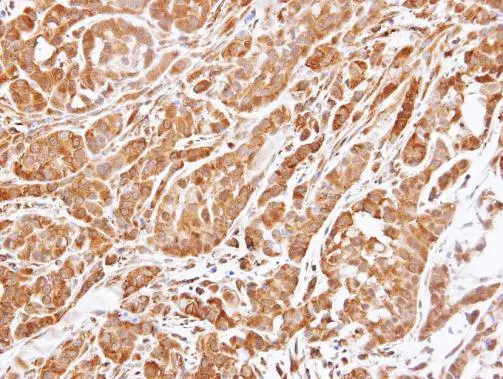
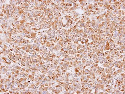
Immunohistochemical analysis of paraffin-embedded A549 xenograft , using 14-3-3 epsilon(GTX109090) antibody at 1:500 dilution.
Antigen Retrieval: Trilogy? (EDTA based, pH 8.0) buffer, 15min
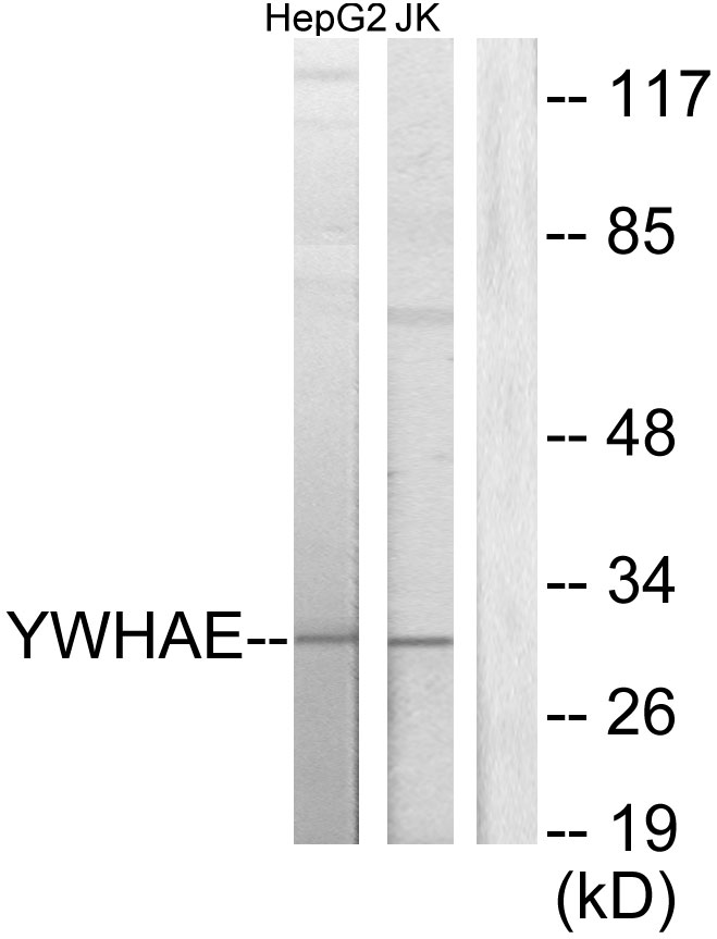
![14-3-3 epsilon antibody [N1C3] immunoprecipitates YWHAE protein in IP experiments. IP samples: HeLa whole cell extract A. 40 μg HeLa whole cell extract B. Control with 4 μg of preimmune Rabbit IgG C. Immunoprecipitation of YWHAE protein by 4 μg 14-3-3 epsilon antibody [N1C3] (GTX109090) 12 % SDS-PAGE The immunoprecipitated YWHAE protein was detected by 14-3-3 epsilon antibody [N1C3] (GTX109090) diluted at 1:1000. [EasyBlot anti-rabbit IgG (GTX221666-01) was used as a secondary reagent]](https://www.genetex.com/upload/website/prouct_img/normal/GTX109090/GTX109090_40016_IP_w_23060120_900.webp "14-3-3 epsilon antibody [N1C3] immunoprecipitates YWHAE protein in IP experiments. IP samples: HeLa whole cell extract A. 40 μg HeLa whole cell extract B. Control with 4 μg of preimmune Rabbit IgG C. Immunoprecipitation of YWHAE protein by 4 μg 14-3-3 epsilon antibody [N1C3] (GTX109090) 12 % SDS-PAGE The immunoprecipitated YWHAE protein was detected by 14-3-3 epsilon antibody [N1C3] (GTX109090) diluted at 1:1000. [EasyBlot anti-rabbit IgG (GTX221666-01) was used as a secondary reagent]")
 antibody at 1:200 dilution.")
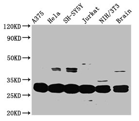
![Various whole cell extracts (30 μg) were separated by 12% SDS-PAGE, and the membrane was blotted with 14-3-3 epsilon antibody [N1C3] (GTX109090) diluted at 1:5000. The HRP-conjugated anti-rabbit IgG antibody (GTX213110-01) was used to detect the primary antibody.](https://www.genetex.com/upload/website/prouct_img/normal/GTX109090/GTX109090_43811_20200103_WB_24110700_897.webp "Various whole cell extracts (30 μg) were separated by 12% SDS-PAGE, and the membrane was blotted with 14-3-3 epsilon antibody [N1C3] (GTX109090) diluted at 1:5000. The HRP-conjugated anti-rabbit IgG antibody (GTX213110-01) was used to detect the primary antibody.")
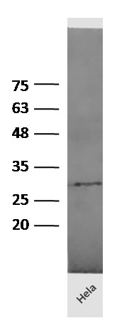
![Mouse tissue extract (50 μg) was separated by 12% SDS-PAGE, and the membrane was blotted with 14-3-3 epsilon antibody [N1C3] (GTX109090) diluted at 1:10000. The HRP-conjugated anti-rabbit IgG antibody (GTX213110-01) was used to detect the primary antibody.](https://www.genetex.com/upload/website/prouct_img/normal/GTX109090/GTX109090_43811_20200103_WB_M_brain_24110700_266.webp "Mouse tissue extract (50 μg) was separated by 12% SDS-PAGE, and the membrane was blotted with 14-3-3 epsilon antibody [N1C3] (GTX109090) diluted at 1:10000. The HRP-conjugated anti-rabbit IgG antibody (GTX213110-01) was used to detect the primary antibody.")
Immunohistochemical analysis of paraffin-embedded A549 xenograft , using 14-3-3 epsilon(GTX109090) antibody at 1:500 dilution.
Antigen Retrieval: Trilogy? (EDTA based, pH 8.0) buffer, 15min
14-3-3 epsilon antibody [N1C3]
GTX109090
ApplicationsImmunoFluorescence, ImmunoPrecipitation, Western Blot, ImmunoCytoChemistry, ImmunoHistoChemistry, ImmunoHistoChemistry Paraffin
Product group Antibodies
ReactivityHuman, Mouse
TargetYWHAE
Overview
- SupplierGeneTex
- Product Name14-3-3 epsilon antibody [N1C3]
- Delivery Days Customer9
- Application Supplier NoteWB: 1:1000-1:10000. ICC/IF: 1:100-1:1000. IHC-P: 1:100-1:1000. IP: 1:100-1:500. *Optimal dilutions/concentrations should be determined by the researcher.Not tested in other applications.
- ApplicationsImmunoFluorescence, ImmunoPrecipitation, Western Blot, ImmunoCytoChemistry, ImmunoHistoChemistry, ImmunoHistoChemistry Paraffin
- CertificationResearch Use Only
- ClonalityPolyclonal
- Concentration1.3 mg/ml
- ConjugateUnconjugated
- Gene ID7531
- Target nameYWHAE
- Target descriptiontyrosine 3-monooxygenase/tryptophan 5-monooxygenase activation protein epsilon
- Target synonyms14-3-3E, HEL2, KCIP-1, MDCR, MDS, 14-3-3 protein epsilon, 14-3-3 epsilon, epididymis luminal protein 2, mitochondrial import stimulation factor L subunit, protein kinase C inhibitor protein-1, tyrosine 3-monooxygenase/tryptophan 5-monooxygenase activation protein, epsilon polypeptide, tyrosine 3/tryptophan 5 -monooxygenase activation protein, epsilon polypeptide
- HostRabbit
- IsotypeIgG
- Protein IDP62258
- Protein Name14-3-3 protein epsilon
- Scientific DescriptionThis gene product belongs to the 14-3-3 family of proteins which mediate signal transduction by binding to phosphoserine-containing proteins. This highly conserved protein family is found in both plants and mammals, and this protein is 100% identical to the mouse ortholog. It interacts with CDC25 phosphatases, RAF1 and IRS1 proteins, suggesting its role in diverse biochemical activities related to signal transduction, such as cell division and regulation of insulin sensitivity. It has also been implicated in the pathogenesis of small cell lung cancer. Two transcript variants, one protein-coding and the other non-protein-coding, have been found for this gene. [provided by RefSeq]
- ReactivityHuman, Mouse
- Storage Instruction-20°C or -80°C,2°C to 8°C
- UNSPSC41116161
Datasheet
Related products
Product group Antibodies
ApplicationsWestern Blot, ELISA
ReactivityHuman, Mouse, Rat
- SizePrice
Product group Antibodies
Anti-YWHAE Antibody144-01058
ApplicationsWestern Blot
ReactivityHuman, Mouse, Rat
TargetYWHAE
- SizePrice

Product group Antibodies
Anti-YWHAE Antibody Picoband(r)A01687-4-CARRIER-FREE
ApplicationsFlow Cytometry, ImmunoFluorescence, Western Blot, ELISA, ImmunoCytoChemistry, ImmunoHistoChemistry
ReactivityHuman, Mouse, Rat
TargetYWHAE
- SizePrice
Product group Antibodies
ApplicationsImmunoFluorescence, Western Blot, ELISA, ImmunoCytoChemistry, ImmunoHistoChemistry, ImmunoHistoChemistry Frozen, ImmunoHistoChemistry Paraffin
ReactivityBovine, Chicken, Human, Mouse, Rabbit, Rat, Sheep
TargetYWHAE
- SizePrice
Product group Antibodies
YWHAE Polyclonal AntibodyCAC14002
ApplicationsImmunoFluorescence, Western Blot, ELISA, ImmunoHistoChemistry
ReactivityMouse
TargetYWHAE
- SizePrice
Product group Antibodies
YWHAE AntibodyCSB-PA026287DA01HU
ApplicationsImmunoFluorescence, Western Blot, ELISA, ImmunoHistoChemistry
ReactivityHuman, Mouse
TargetYWHAE
- SizePrice

Product group Antibodies
Anti-YWHAE AntibodyHPA008445
ApplicationsWestern Blot, ImmunoHistoChemistry
ReactivityHuman, Mouse, Rat
TargetYWHAE
- SizePrice
Product group Antibodies
14-3-3 epsilon antibody [N2C3]GTX113576
ApplicationsImmunoFluorescence, Western Blot, ImmunoCytoChemistry, ImmunoHistoChemistry, ImmunoHistoChemistry Paraffin
ReactivityHuman, Mouse
TargetYWHAE
- SizePrice
Product group Antibodies
14-3-3 epsilon antibodyGTX55485
ApplicationsImmunoFluorescence, Western Blot, ImmunoCytoChemistry
ReactivityHuman, Mouse, Rat
TargetYWHAE
- SizePrice
Product group Antibodies
14-3-3 epsilon antibody [AT4F8]GTX57563
ApplicationsImmunoFluorescence, Western Blot, ImmunoCytoChemistry
ReactivityHuman, Mouse
TargetYWHAE
- SizePrice