Bio-Connect
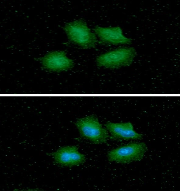
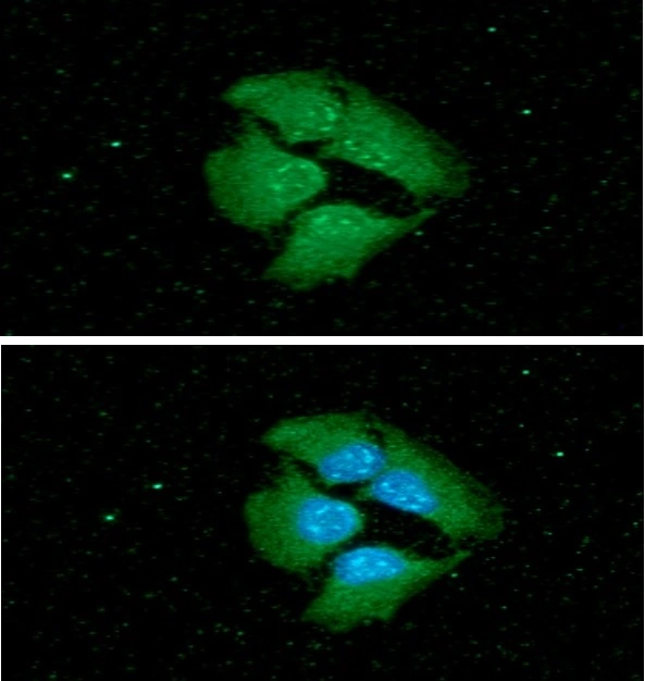
ICC/IF analysis of HeLa cells using GTX57563 14-3-3 epsilon antibody. Blue: DAPI Green: Primary antibody Dilution: 1:100

14-3-3 epsilon antibody [AT4F8]

GTX57563
GeneTex
ApplicationsImmunoFluorescence, Western Blot, ImmunoCytoChemistry
Product group Antibodies
TargetYWHAE
Sign in to order and to see your custom pricing.
Large volume orders?
Order with a bulk request

Overview

  • Supplier
    GeneTex
  • Product Name
    14-3-3 epsilon antibody [AT4F8]
  • Delivery Days Customer
    9
  • Applications
    ImmunoFluorescence, Western Blot, ImmunoCytoChemistry
  • Certification
    Research Use Only
  • Clonality
    Monoclonal
  • Clone ID
    AT4F8
  • Concentration
    1 mg/ml
  • Conjugate
    Unconjugated
  • Gene ID7531
  • Target name
    YWHAE
  • Target description
    tyrosine 3-monooxygenase/tryptophan 5-monooxygenase activation protein epsilon
  • Target synonyms
    14-3-3 epsilon; 14-3-3 protein epsilon; 14-3-3E; epididymis luminal protein 2; HEL2; KCIP-1; MDCR; MDS; mitochondrial import stimulation factor L subunit; protein kinase C inhibitor protein-1; tyrosine 3/tryptophan 5 -monooxygenase activation protein, epsilon polypeptide; tyrosine 3-monooxygenase/tryptophan 5-monooxygenase activation protein, epsilon polypeptide
  • Host
    Mouse
  • Isotype
    IgG2b
  • Scientific Description
    This gene product belongs to the 14-3-3 family of proteins which mediate signal transduction by binding to phosphoserine-containing proteins. This highly conserved protein family is found in both plants and mammals, and this protein is 100% identical to the mouse ortholog. It interacts with CDC25 phosphatases, RAF1 and IRS1 proteins, suggesting its role in diverse biochemical activities related to signal transduction, such as cell division and regulation of insulin sensitivity. It has also been implicated in the pathogenesis of small cell lung cancer. Two transcript variants, one protein-coding and the other non-protein-coding, have been found for this gene. [provided by RefSeq, Aug 2008]
  • Storage Instruction
    -20°C or -80°C,2°C to 8°C
  • UNSPSC
    12352203

Related products

Product group Antibodies
Biomatik
ApplicationsImmunoFluorescence, Western Blot, ELISA, ImmunoHistoChemistry
TargetYWHAE
  • SizePrice
Product group Antibodies
Assay Genie
TargetYWHAE
  • SizePrice
Product group Antibodies
References
GeneTex
ApplicationsImmunoFluorescence, ImmunoPrecipitation, Western Blot, ImmunoCytoChemistry, ImmunoHistoChemistry, ImmunoHistoChemistry Paraffin
TargetYWHAE
  • SizePrice
Product group Antibodies
GeneTex
ApplicationsImmunoFluorescence, Western Blot, ImmunoCytoChemistry, ImmunoHistoChemistry, ImmunoHistoChemistry Paraffin
TargetYWHAE
  • SizePrice
Product group Antibodies
MedChem Express
TargetYWHAE
  • SizePrice
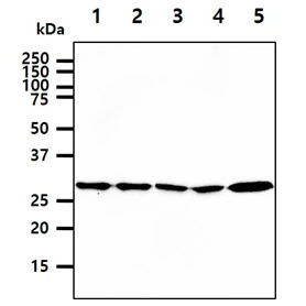
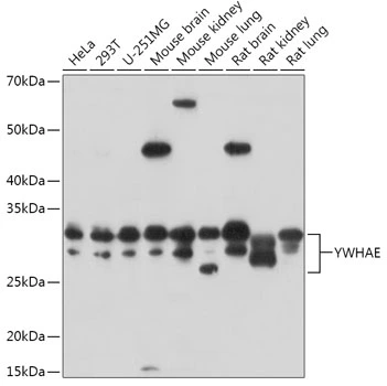
WB analysis of various sample lysates using GTX55485 14-3-3 epsilon antibody. Dilution : 1:3000 Loading : 25microg per lane
Product group Antibodies
GeneTex
ApplicationsImmunoFluorescence, Western Blot, ImmunoCytoChemistry
TargetYWHAE
  • SizePrice
Immunohistochemical staining of human cerebral cortex shows strong nuclear and cytoplasmic positivity in neuronal cells.
Product group Antibodies
Atlas Antibodies
ApplicationsWestern Blot, ImmunoHistoChemistry
TargetYWHAE
  • SizePrice
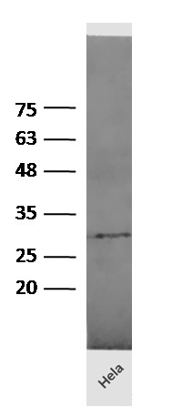
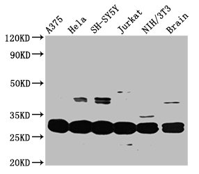
Western Blot Positive WB detected in: A375 whole cell lysate, Hela whole cell lysate, SH-SY5Y whole cell lysate, Jurkat whole cell lysate, NIH/3T3 whole cell lysate, Mouse brain tissue All lanes: YWHAE antibody at 3.3microg/ml Secondary Goat polyclonal to rabbit IgG at 1/50000 dilution Predicted band size: 30, 27 kDa Observed band size: 30 kDa
Product group Antibodies
Cusabio
YWHAE AntibodyCSB-PA026287DA01HU
ApplicationsImmunoFluorescence, Western Blot, ELISA, ImmunoHistoChemistry
TargetYWHAE
  • SizePrice
Figure 1. Western blot analysis of YWHAE using anti-YWHAE antibody (A01687-4). Electrophoresis was performed on a 5-20% SDS-PAGE gel at 70V (Stacking gel) / 90V (Resolving gel) for 2-3 hours. The sample well of each lane was loaded with 50ug of sample under reducing conditions. Lane 1: human Hela whole cell lysates, Lane 2: human Jurkat whole cell lysates, Lane 3: human HepG2 whole cell lysates, Lane 4: human SH-SY5Y whole cell lysates, Lane 5: human HEK293 whole cell lysates, Lane 6: human SW620 whole cell lysates, Lane 7: human A549 whole cell lysates, Lane 8: human Raji whole cell lysates, Lane 9: rat brain tissue lysates, Lane 10: mouse brain tissue lysates, Lane 11: mouse NIH/3T3 whole cell lysates, Lane 12: mouse RAW264.7 whole cell lysates. After Electrophoresis, proteins were transferred to a Nitrocellulose membrane at 150mA for 50-90 minutes. Blocked the membrane with 5% Non-fat Milk/ TBS for 1.5 hour at RT. The membrane was incubated with rabbit anti-YWHAE antigen affinity purified polyclonal antibody (Catalog # A01687-4) at 0.5 microg/mL overnight at 4°C, then washed with TBS-0.1%Tween 3 times with 5 minutes each and probed with a goat anti-rabbit IgG-HRP secondary antibody at a dilution of 1:10000 for 1.5 hour at RT. The signal is developed using an Enhanced Chemiluminescent detection (ECL) kit (Catalog # EK1002) with Tanon 5200 system. A specific band was detected for YWHAE at approximately 28KD. The expected band size for YWHAE is at 28KD.
Product group Antibodies
Boster Bio
Anti-YWHAE Antibody Picoband(r)A01687-4-CARRIER-FREE
ApplicationsFlow Cytometry, ImmunoFluorescence, Western Blot, ELISA, ImmunoCytoChemistry, ImmunoHistoChemistry
TargetYWHAE
  • SizePrice
Anti-14-3-3 epsilon Antibody
Product group Antibodies
Antibodies.com
ApplicationsWestern Blot, ELISA
  • SizePrice