

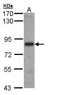
Sample (30 ug of whole cell lysate) A: Hep G2 (GTX27900) 7.5% SDS PAGE GTX109708 diluted at 1:1000
Sample (30 ug of whole cell lysate) A: Hep G2 (GTX27900) 7.5% SDS PAGE GTX109708 diluted at 1:1000
3BP2 antibody
GTX109708
ApplicationsWestern Blot
Product group Antibodies
ReactivityHuman
TargetSH3BP2
Overview
- SupplierGeneTex
- Product Name3BP2 antibody
- Delivery Days Customer9
- Application Supplier NoteWB: 1:500-1:3000. *Optimal dilutions/concentrations should be determined by the researcher.Not tested in other applications.
- ApplicationsWestern Blot
- CertificationResearch Use Only
- ClonalityPolyclonal
- Concentration1 mg/ml
- ConjugateUnconjugated
- Gene ID6452
- Target nameSH3BP2
- Target descriptionSH3 domain binding protein 2
- Target synonyms3BP-2, 3BP2, CRBM, CRPM, RES4-23, SH3 domain-binding protein 2, Abl-SH3 binding protein 2, TNFAIP3 interacting protein 2, epididymis secretory sperm binding protein
- HostRabbit
- IsotypeIgG
- Protein IDP78314
- Protein NameSH3 domain-binding protein 2
- Scientific DescriptionThe protein encoded by this gene has an N-terminal pleckstrin homology (PH) domain, an SH3-binding proline-rich region, and a C-terminal SH2 domain. The protein binds to the SH3 domains of several proteins including the ABL1 and SYK protein tyrosine kinases , and functions as a cytoplasmic adaptor protein to positively regulate transcriptional activity in T, natural killer (NK), and basophilic cells. Mutations in this gene result in cherubism. Multiple transcript variants encoding different isoforms have been found for this gene. [provided by RefSeq]
- ReactivityHuman
- Storage Instruction-20°C or -80°C,2°C to 8°C
- UNSPSC41116161
Datasheet
Related products
Product group Antibodies
SH3BP2 AntibodyCSB-PA021224LA01HU
ApplicationsELISA, ImmunoHistoChemistry
ReactivityHuman
TargetSH3BP2
- SizePrice
Product group Antibodies
Anti-SH3BP2 AntibodyA12856
ApplicationsWestern Blot
ReactivityHuman, Mouse, Rat
- SizePrice
Product group Antibodies
Anti-SH3BP2 Antibody144-61794
ApplicationsWestern Blot
ReactivityHuman, Mouse, Rat
TargetSH3BP2
- SizePrice

Product group Antibodies
Anti-SH3BP2 AntibodyHPA036789
ApplicationsImmunoCytoChemistry
ReactivityHuman
TargetSH3BP2
- SizePrice

Product group Antibodies
Anti-3BP2/SH3BP2 Antibody Picoband(r)A04008-1-CARRIER-FREE
ApplicationsFlow Cytometry, Western Blot, ELISA
ReactivityHuman
TargetSH3BP2
- SizePrice
Product group Antibodies

SH3BP2 AntibodyLS-C496951
ApplicationsWestern Blot
ReactivityHuman, Mouse, Rat
TargetSH3BP2
- SizePrice